Table Top and Unit arm included. If you need an electric table mount, let us know.
Used Haag Streit AT900 Tonometer
Power Cable
Instruction Manual.
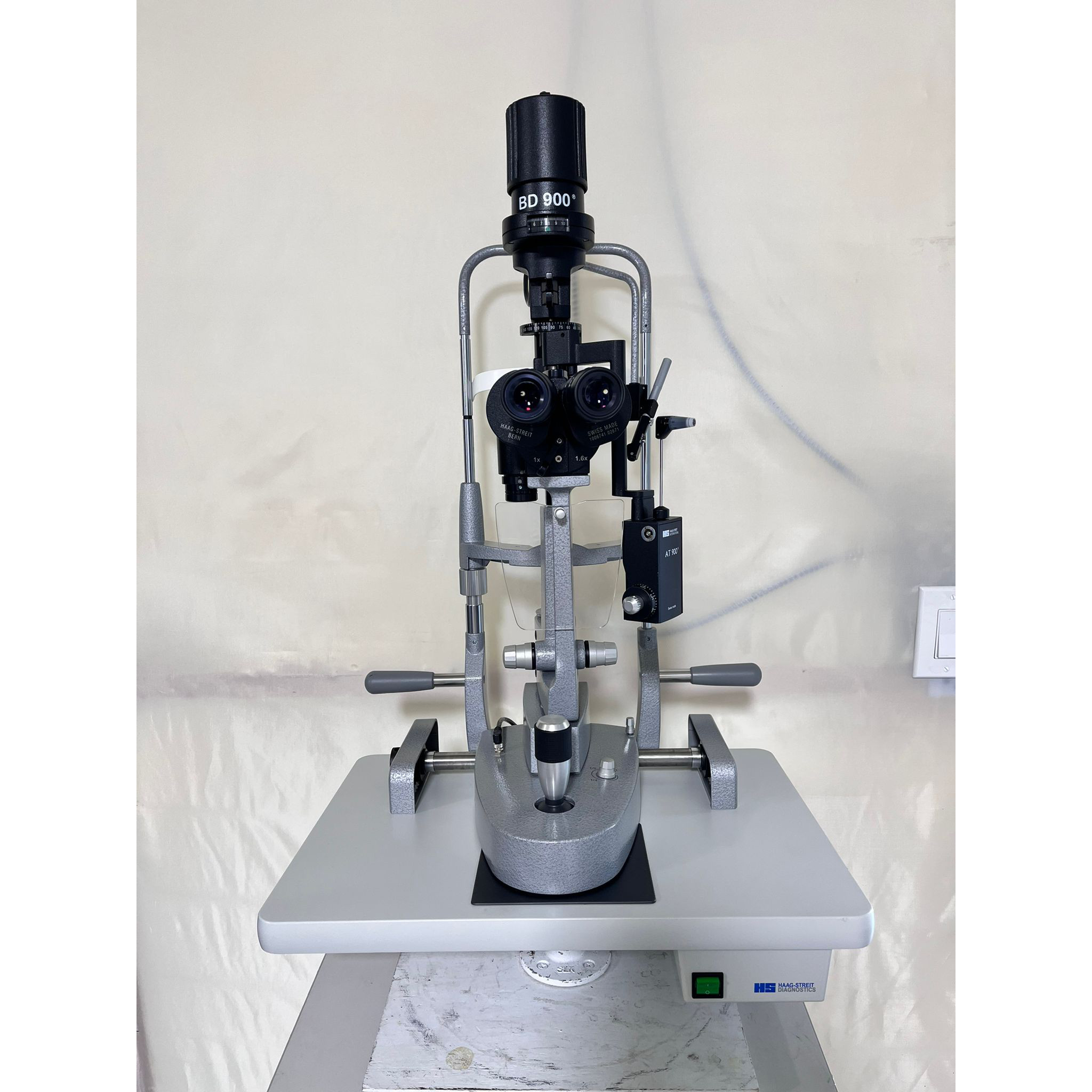
Detail Haag Streit BD900
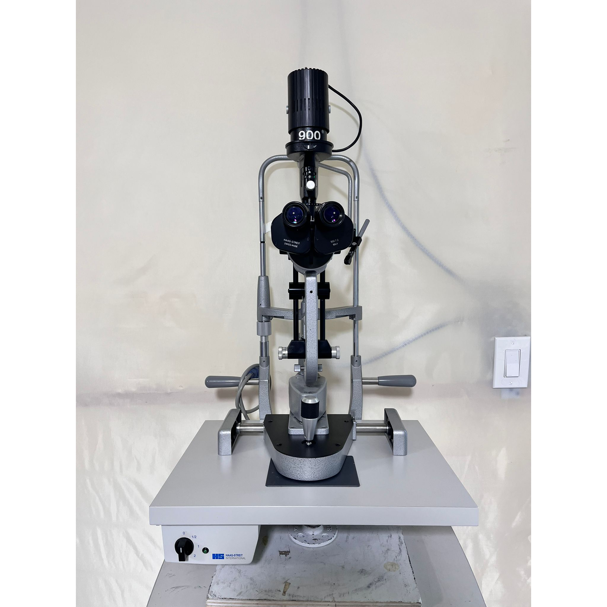
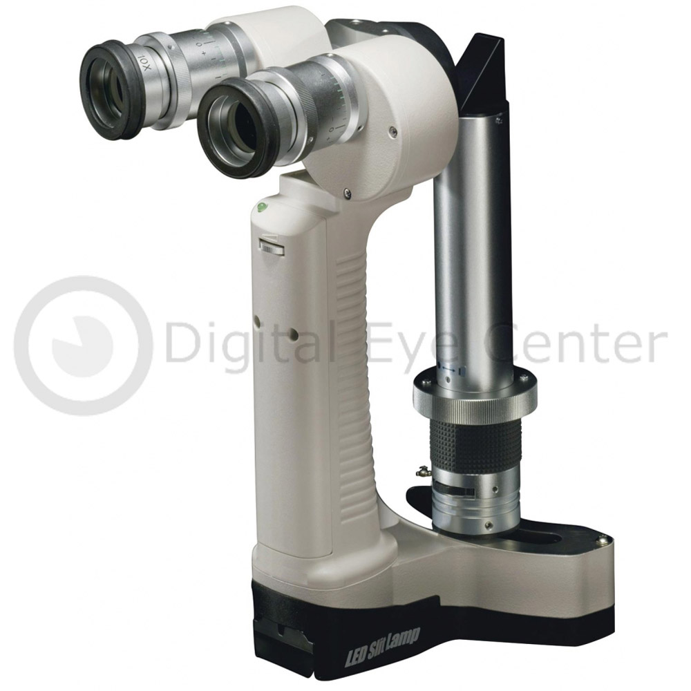
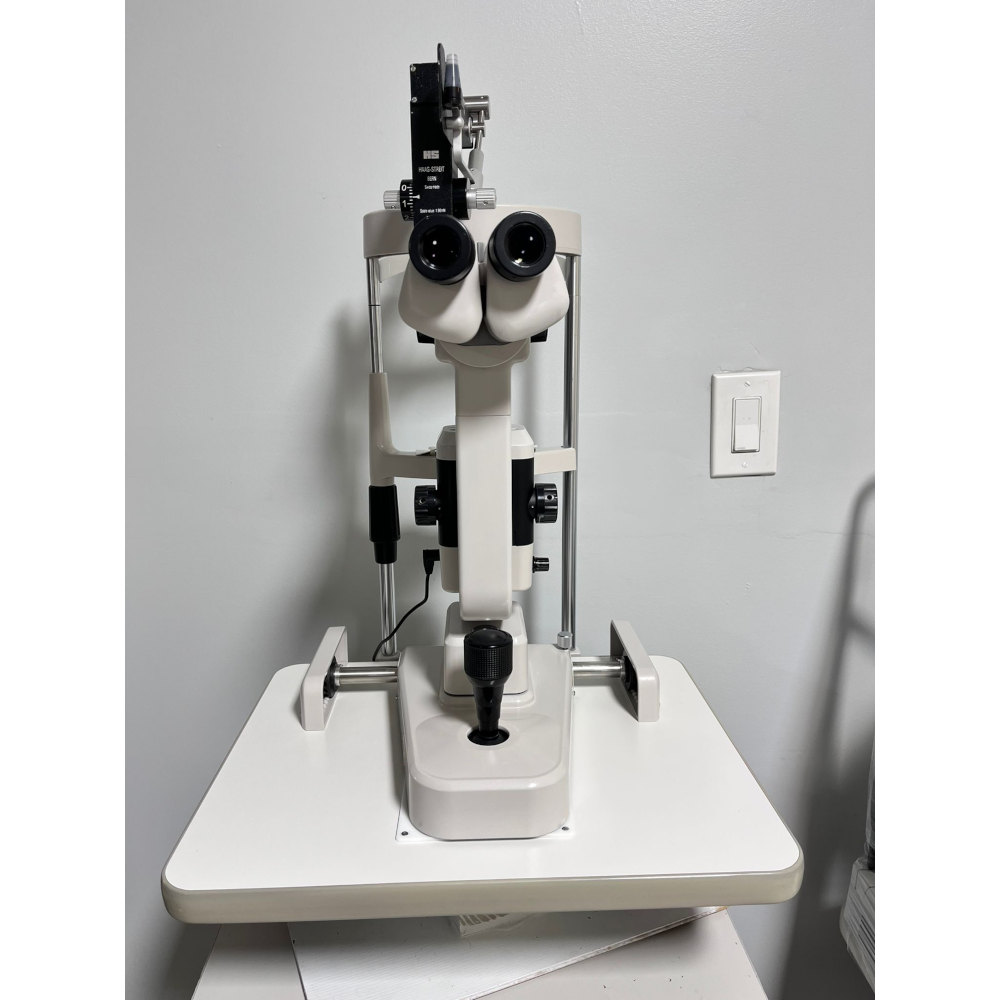
The preowned Haag-Streit BD 900 Slit Lamp is a versatile slit lamp specialized for examination of the anterior segment, and it is ideal for use in either an ophthalmologist’s exam room or the emergency room. The BD 900 is capable of 10- and 16-times magnification and has a built-in recording video port. It is especially suited for contact lens fitting.
Features
Maintained essential qualities of BM 900 and BQ 900
Table Top and Unit arm included. If you need an electric table mount, let us know.
Power Cable
Instruction Manual.
Description Haag-Streit BQ 900 LED
Excellent optics
The quality of the optical system determines the results of whatever application a slit lamp is used for. The BQ 900 has an elaborate optical system manufactured to meet the highest quality requirements. The result is a superb view, allowing accurate diagnostics, safe patient treatment, and stunning imaging results.
Superior mechanics
Haag-Streit has stood for high-precision mechanics since its foundation more than 160 years ago. This experience, outstanding Swiss engineering, and high-grade materials ensure perfect mechanics in the BQ 900, which can last decades.
Table Top and Unit arm included. If you need an electric table mount, let us know.
Power Cable
Instruction Manual.
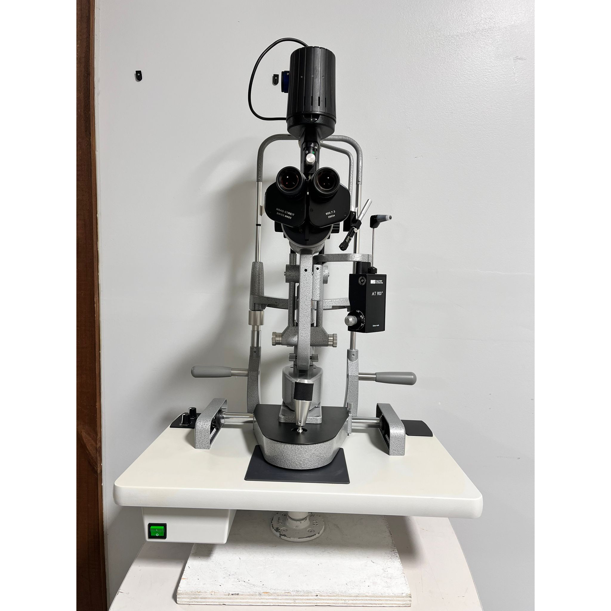
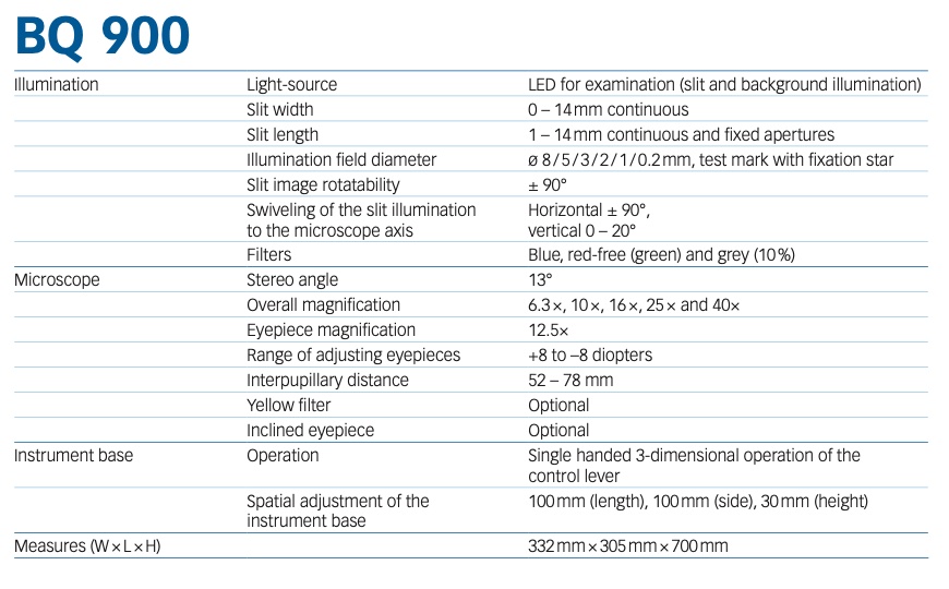
Description Haag-Streit BQ 900
The Haag-Streit pre-owned BQ 900 Slit Lamp is a versatile slit lamp specialized for examination of the anterior segment, and it is ideal for use in either an ophthalmologist’s exam room or the emergency room. The BQ-900 magnifies from 6.3 × up to 40 × selectable in 5 fixed steps.
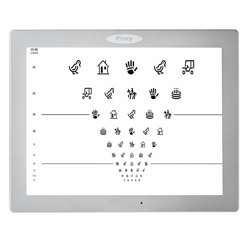
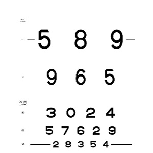
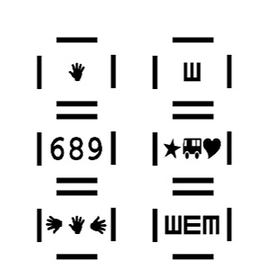
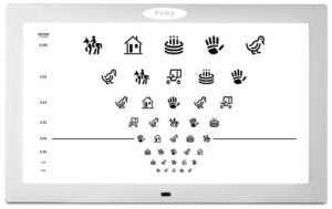
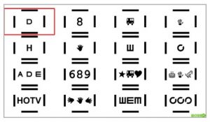
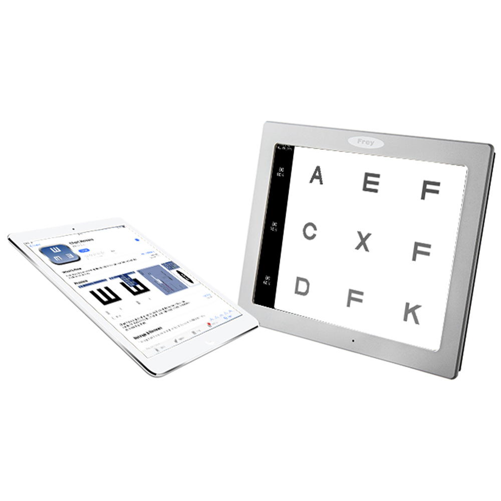
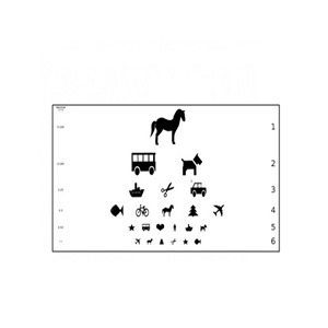
Multiple test charts are available including letters charts, number charts, symbol charts.
The Frey CP-400 Chart Panel provides multiple chart display models and scales to allow the user to select the best method for the test procedure.
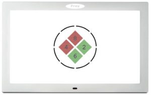
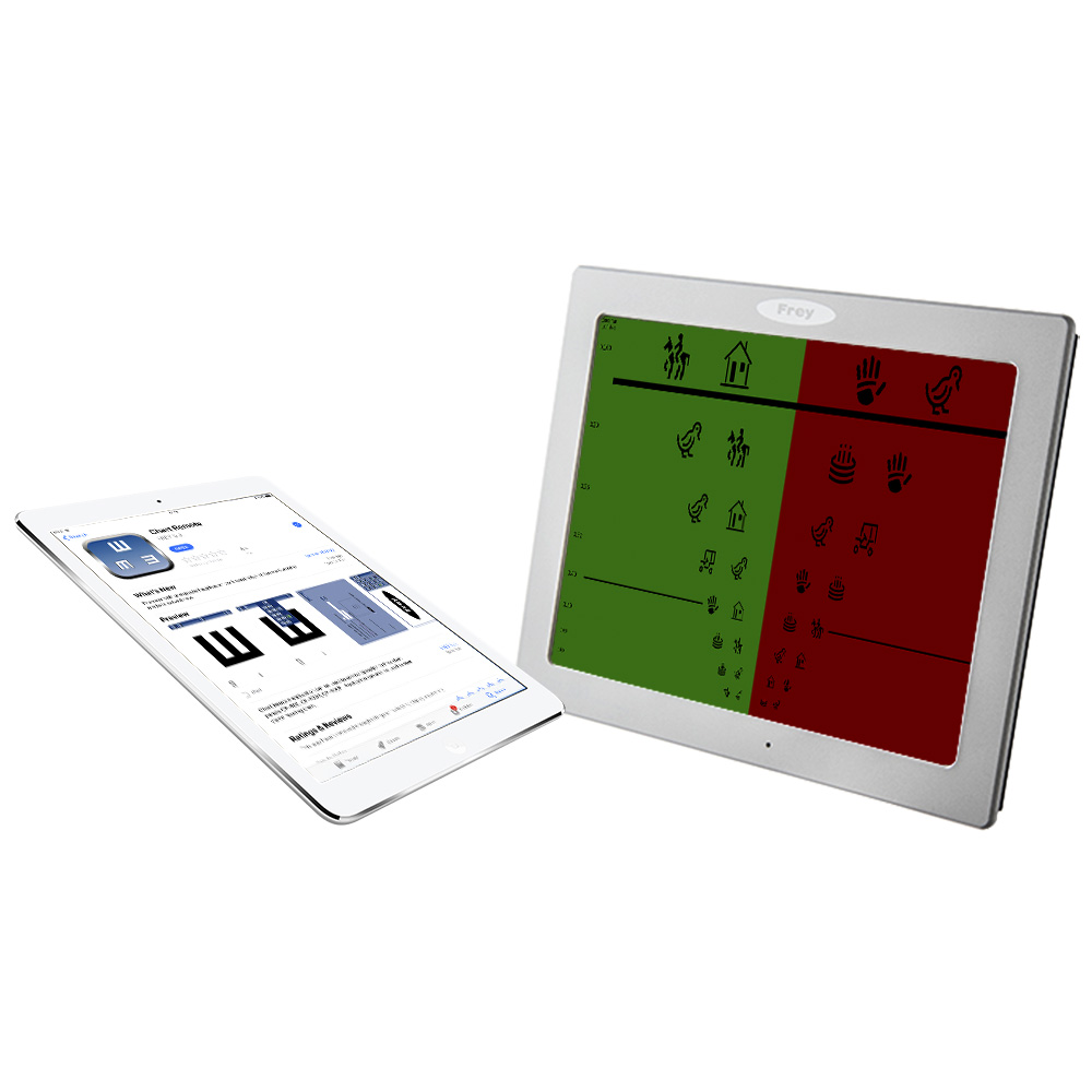
Color Vision testing
The CP-400 has built-in color vision charts allowing to examine patient’s color recognition accuracy.
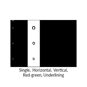
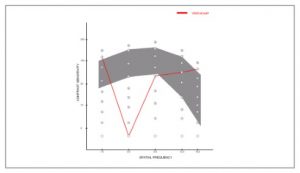
Contrast Sensitivity Testing
Contrast testing can be performed on CP-400 using optotypes presented at different contrast level or for our more demanding customers sinusoidal bar grading test.
Patient Education Module
The patient education module of CP-400 Chart Panel provides the user with storage for educational pictures and videos which can be used for explaining eye disorders to the patient.
Test Reports
With the assistance of the remote control the user can record results of the tests. At the end of the test a report can be created and stored in the device memory in .pdf format. The report can be transferred to an external computer with the use of USB memory stick or a WLAN connection.
User defined test sequences
For users willing to standardize their test procedures the CP-400 provides programming functions. Up to 3 programs can be defined by the user and then started with just one click on the remote .
Automated phoropter communication
The CP-400 Chart panel can communicate wirelessly with different models of auto phoropters. This feature allows users to extend the functionality of their existing phoropter systems .
Wireless connectivity option
Wireless communication is a feature available with optional USB WiFi module. It allows to communicate with CP-400 chart panel in computer network and access test reports and educational materials stored on the device.
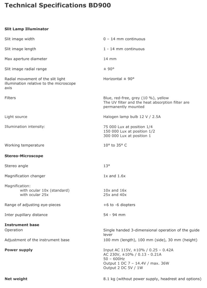
Technical Specifications
Display size
24″ diagonal
Dimensions
605 x 370 x 30 mm (L/W/H)
Weight
3,5 kg
Power supply
External INPUT: 100 – 240 ~0.9A 50/60 Hz. OUTPUT: 12.0V 3.0A DC
Power consumption
35 W max
Picture to Picture change time
0.5s
Refraction distance
2.9 to 6.1m
Background luminance
200 cd/m2
Auto off function
5, 10, 15 min – adjustable
Number of user test programs
3 programs, 15 steps each
VESA mount hole pattern
200 x 100mm
Software Features
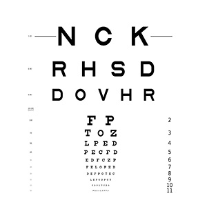
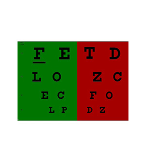
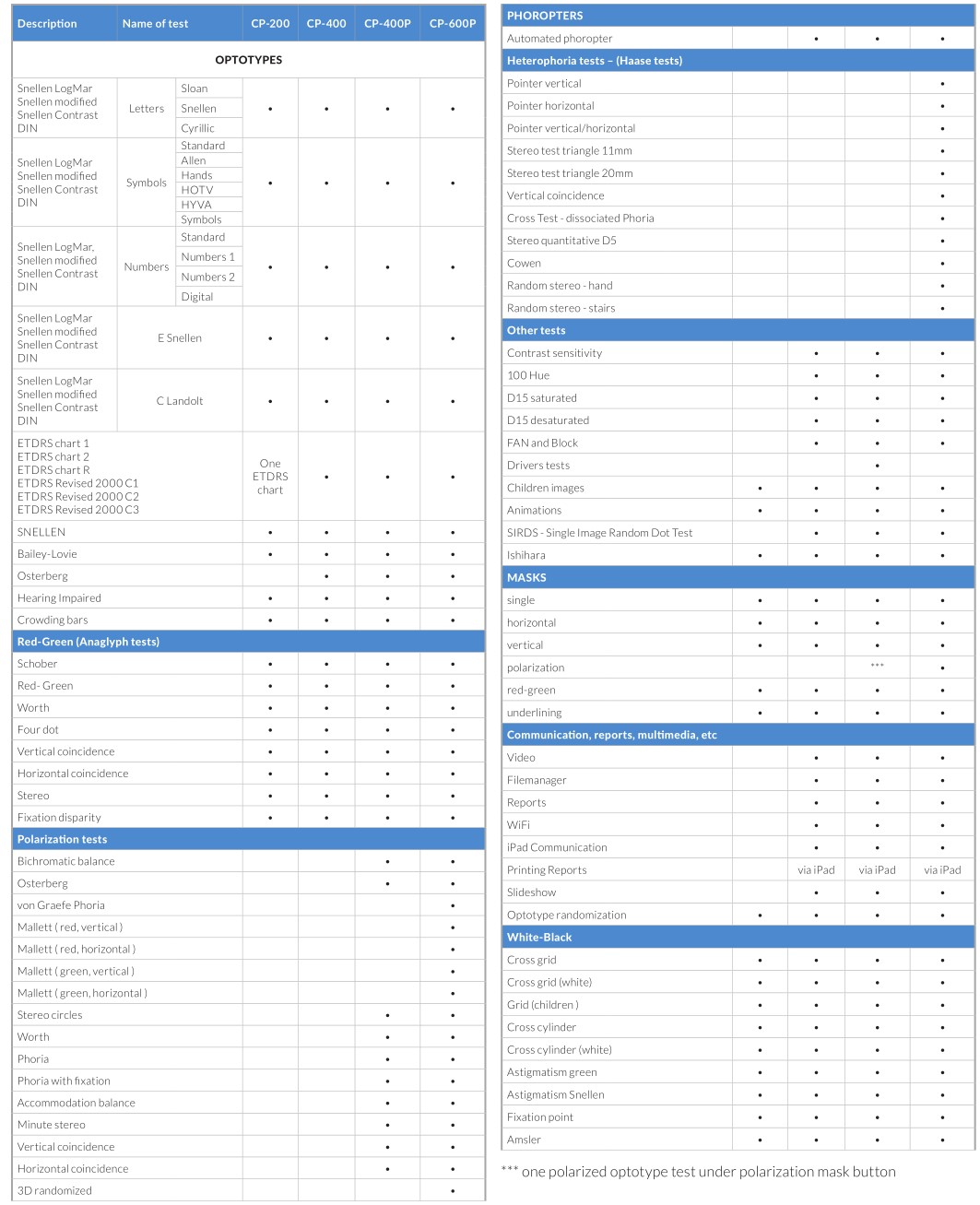
Letter Charts
Available optotypes: Sloan, Snellen, Cyrillic
Presentation modes: Snellen, LogMar, Snellen modified, Snellen contrast, DIN
Symbol Charts
Available optotypes: Standard, Allen, Hands, HOTV, HYVA, Symbols
Presentation modes: Snellen, LogMar, Snellen modified, Snellen contrast, DIN
Number Charts
Available optotypes:
Standard, Numbers 1, Numbers 2, Digital
Presentation modes:
Snellen, LogMar, Snellen modified, Snellen contrast, DIN
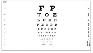
Snellen Charts
Presentation modes:
Snellen, LogMar, Snellen modified, Snellen contrast, DIN
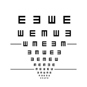
Landolt Charts
Presentation modes:
Snellen, LogMar, Snellen modified, Snellen contrast, DIN
The Frey Chart Panel suite of devices is one the world’s leading fully featured digital acuity system with more than 5000 devices installed worldwide.
Presented in a slim line and attractive LCD panel with our proprietary built-in powerful computer, Frey Chart Panels enhances the look of any modern clinical practice designed to perform a comprehensive suite of recognized international visual acuity tests supported by easily customized reporting tools to suit clinician or practice needs.
Clinical advantages of the Frey Chart Panel technology
Fully integrated customized design designed to eliminate the risk of any computer related software or hardware incompatibility.
Extensive range of tests and functions that result in a precise determination of ocular diseases and common medical conditions.
Extensive range of tests and Optotypes with upgrade capability.
Complete range of contrast, color and stereo tests.
Patient education tools.
Comprehensive Optotype range including EU,UK, US and International standards.
Precise adjustment of working distance and Optotype.
Smooth and silent operation.
Optimized for illuminated rooms.
Built-in proprietary computer.
Test reporting.
Speech and hearing impaired tests.
Animations and videos.
Random and User programmable sequencing.
Mirrored setup.
Continuous technology upgrades.
Test Report customized Patient Education and Video Functions.
Slim Line space saving design, easily mounted
Ease of Use
Frey Chart Panels can be mounted directly on the wall or with use of VESA Standard adjustable Wall Mount or Desk Stand.
A simple to use IR remote with direct commands is intuitive and simple to use. Tests can be carried out in daylight illumination conditions creating a comfortable and convenient environment for patients and clinician.
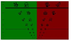
Enhanced Children Tests & Fixation Methods
A complete suite of Optotypes, including HOTV and ALLEN Pre-School tests are supported by Animations, Colorful Pictures and Videos.
Superior Contrast Testing
Threshold Optotype Testing: Complements and extends the assessment in visual acuity function.
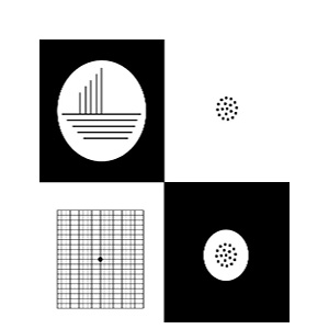
Spatial Frequency Testing: A proprietary contrast test for the early diagnosis of Cataract, Diabetic Retinopathy and other ocular diseases.
Test Result Reporting
Patient details can be easily entered into the system upon which the clinician can perform a sequence of acuity and visual tests. Test results can be stored in the Frey Chart Panel memory.
Low Vision AMD Optotypes
Standardized vision tests for patients with Age-related Macular Degeneration (AMD)
with Optotypes size and contrast alteration at a calibrated distance.
Multiple test charts are available including letters charts, number charts, symbol charts.
The Frey CP-400 Chart Panel provides multiple chart display models and scales to allow the user to select the best method for the test procedure.
Color Vision testing
The CP-400 has built-in color vision charts allowing to examine patient’s color recognition accuracy.
Contrast Sensitivity Testing
Contrast testing can be performed on CP-400 using optotypes presented at different contrast level or for our more demanding customers sinusoidal bar grading test.
Polarized Stereo Tests
Polarization filter technology used in model CP-400P assures high separation between patient visual channels, combined with reach selection of vision acuity test charts makes CP-400P ideal tool for users searching for advanced acuity and vision system condition test device.
Patient Education Module
The patient education module of CP-400 Chart Panel provides the user with storage for educational pictures and videos which can be used for explaining eye disorders to the patient.
Test Reports
With the assistance of the remote control the user can record results of the tests. At the end of the test a report can be created and stored in the device memory in .pdf format. The report can be transferred to an external computer with the use of USB memory stick or a WLAN connection.
User defined test sequences
For users willing to standardize their test procedures the CP-400 provides programming functions. Up to 3 programs can be defined by the user and then started with just one click on the remote .
Automated phoropter communication
The CP-400 Chart panel can communicate wirelessly with different models of auto phoropters. This feature allows users to extend the functionality of their existing phoropter systems .
Wireless connectivity option
Wireless communication is a feature available with optional USB WiFi module. It allows to communicate with CP-400 chart panel in computer network and access test reports and educational materials stored on the device.
Technical Specifications
Display size
23″ diagonal
Dimensions
583 x 359 x 30mm (L/W/H)
Weight
3,9 kg
Power supply
External INPUT: 100 – 240 ~1.5A 50/60 Hz. OUTPUT: 12.0V 5.0A DC
Power consumption
60 W max
Picture to Picture change time
0.5s
Refraction distance
2.9 to 6.1m
Background luminance
200 cd/m2
Auto off function
5, 10, 15 min – adjustable
Number of user test programs
3 programs, 15 steps each
VESA mount hole pattern
200 x 100mm
Software Features
Letter Charts
Available optotypes: Sloan, Snellen, Cyrillic
Presentation modes: Snellen, LogMar, Snellen modified, Snellen contrast, DIN
Symbol Charts
Available optotypes: Standard, Allen, Hands, HOTV, HYVA, Symbols
Presentation modes: Snellen, LogMar, Snellen modified, Snellen contrast, DIN
Number Charts
Available optotypes:
Standard, Numbers 1, Numbers 2, Digital
Presentation modes:
Snellen, LogMar, Snellen modified, Snellen contrast, DIN
Snellen Charts
Presentation modes:
Snellen, LogMar, Snellen modified, Snellen contrast, DIN
Landolt Charts
Presentation modes:
Snellen, LogMar, Snellen modified, Snellen contrast, DIN
Our Frey Chart Panel CP-400P can communicate wirelessly with most of the brands of automated phoropters in the market
. This feature allows users to extend the functionality of their existing phoropter systems .
Bailey-Lovie chart
Hearing Impaired
Crowding Bars
Communication, reports, multimedia
Video
File manager
Reports
WiFi
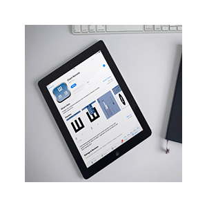
iPad communication (Frey Chart Panel Application)
Printing reports
(cia iPad)
Slideshow
Optotype randomization
Other Tests
FAN and Block
Children images
Animations
SIRDS – Single Image Random Dot Test
Contrast sensitivity
100 Hue
D15 saturated
D15 desaturated
Ishihara
Snellen charts
Osterberg Chart
Masks
The Frey Chart Panel suite of devices is one the world’s leading fully featured digital acuity system with more than 5000 devices installed worldwide.
Presented in a slim line and attractive LCD panel with our proprietary built-in powerful computer, Frey Chart Panels enhances the look of any modern clinical practice designed to perform a comprehensive suite of recognized international visual acuity tests supported by easily customized reporting tools to suit clinician or practice needs.
Clinical advantages of the Frey Chart Panel technology
Fully integrated customized design designed to eliminate the risk of any computer related software or hardware incompatibility.
Extensive range of tests and functions that result in a precise determination of ocular diseases and common medical conditions.
Extensive range of tests and Optotypes with upgrade capability.
Complete range of contrast, color and stereo tests.
Patient education tools.
Comprehensive Optotype range including EU,UK, US and International standards.
Precise adjustment of working distance and Optotype.
Smooth and silent operation.
Optimized for illuminated rooms.
Built-in proprietary computer.
Test reporting.
Speech and hearing impaired tests.
Animations and videos.
Random and User programmable sequencing.
Mirrored setup.
Continuous technology upgrades.
Test Report customized Patient Education and Video Functions.
Slim Line space saving design, easily mounted
Ease of Use
Frey Chart Panels can be mounted directly on the wall or with use of VESA Standard adjustable Wall Mount or Desk Stand.
A simple to use IR remote with direct commands is intuitive and simple to use. Tests can be carried out in daylight illumination conditions creating a comfortable and convenient environment for patients and clinician.
Enhanced Children Tests & Fixation Methods
A complete suite of Optotypes, including HOTV and ALLEN Pre-School tests are supported by Animations, Colorful Pictures and Videos.
Superior Contrast Testing
Threshold Optotype Testing: Complements and extends the assessment in visual acuity function.
Spatial Frequency Testing: A proprietary contrast test for the early diagnosis of Cataract, Diabetic Retinopathy and other ocular diseases.
Test Result Reporting
Patient details can be easily entered into the system upon which the clinician can perform a sequence of acuity and visual tests. Test results can be stored in the Frey Chart Panel memory.
Low Vision AMD Optotypes
Standardized vision tests for patients with Age-related Macular Degeneration (AMD)
with Optotypes size and contrast alteration at a calibrated distance.
Multiple test charts are available including letters charts, number charts, symbol charts.
The Frey CP-400 Chart Panel provides multiple chart display models and scales to allow the user to select the best method for the test procedure.
Color Vision testing
The CP-400 has built-in color vision charts allowing to examine patient’s color recognition accuracy.
Contrast Sensitivity Testing
Contrast testing can be performed on CP-400 using optotypes presented at different contrast level or for our more demanding customers sinusoidal bar grading test.
Polarized Stereo Tests
Polarization filter technology used in model CP-400P assures high separation between patient visual channels, combined with reach selection of vision acuity test charts makes CP-400P ideal tool for users searching for advanced acuity and vision system condition test device.
Patient Education Module
The patient education module of CP-400 Chart Panel provides the user with storage for educational pictures and videos which can be used for explaining eye disorders to the patient.
Test Reports
With the assistance of the remote control the user can record results of the tests. At the end of the test a report can be created and stored in the device memory in .pdf format. The report can be transferred to an external computer with the use of USB memory stick or a WLAN connection.
User defined test sequences
For users willing to standardize their test procedures the CP-400 provides programming functions. Up to 3 programs can be defined by the user and then started with just one click on the remote .
Automated phoropter communication
The CP-400 Chart panel can communicate wirelessly with different models of auto phoropters. This feature allows users to extend the functionality of their existing phoropter systems .
Wireless connectivity option
Wireless communication is a feature available with optional USB WiFi module. It allows to communicate with CP-400 chart panel in computer network and access test reports and educational materials stored on the device.
Technical Specifications
Display size
24″ diagonal
Dimensions
605 x 370 x 30mm (L/W/H)
Weight
3,8 kg
Power supply
External INPUT: 100 – 240 ~1.5A 50/60 Hz. OUTPUT: 12.0V 5.0A DC
Power consumption
35 W max
Picture to Picture change time
0.5s
Refraction distance
2.9 to 6.1m
Background luminance
200 cd/m2
Auto off function
5, 10, 15 min – adjustable
Number of user test programs
3 programs, 15 steps each
VESA mount hole pattern
200 x 100mm
Software Features
Letter Charts
Available optotypes: Sloan, Snellen, Cyrillic
Presentation modes: Snellen, LogMar, Snellen modified, Snellen contrast, DIN
Symbol Charts
Available optotypes: Standard, Allen, Hands, HOTV, HYVA, Symbols
Presentation modes: Snellen, LogMar, Snellen modified, Snellen contrast, DIN
Number Charts
Available optotypes:
Standard, Numbers 1, Numbers 2, Digital
Presentation modes:
Snellen, LogMar, Snellen modified, Snellen contrast, DIN
Snellen Charts
Presentation modes:
Snellen, LogMar, Snellen modified, Snellen contrast, DIN
Landolt Charts
Presentation modes:
Snellen, LogMar, Snellen modified, Snellen contrast, DIN
Our Frey Chart Panel CP-400P can communicate wirelessly with most of the brands of automated phoropters in the market
. This feature allows users to extend the functionality of their existing phoropter systems .
Heterophoria Tests (Hasse tests)
Pointer vertical
Pointer horizontal
Pointer vertical/ horizontal
Stereo test triangle 11 mm
Stereo test triangle 20 mm
Vertical coincidence
Cross test – dissociated Phoria
Stereo quantitative D5
Cowen
Random stereo – hand
Random stereo – stairs
Bailey-Lovie chart
Hearing Impaired
Crowding Bars
Communication, reports, multimedia
Video
File manager
Reports
WiFi
iPad communication (Frey Chart Panel Application)
Printing reports
(cia iPad)
Slideshow
Optotype randomization
Other Tests
FAN and Block
Children images
Animations
SIRDS – Single Image Random Dot Test
Contrast sensitivity
100 Hue
D15 saturated
D15 desaturated
Ishihara
Snellen charts
Osterberg Chart
Masks
The Frey Chart Panel suite of devices is one the world’s leading fully featured digital acuity system with more than 5000 devices installed worldwide.
Presented in a slim line and attractive LCD panel with our proprietary built-in powerful computer, Frey Chart Panels enhances the look of any modern clinical practice designed to perform a comprehensive suite of recognized international visual acuity tests supported by easily customized reporting tools to suit clinician or practice needs.
Clinical advantages of the Frey Chart Panel technology
Fully integrated customized design designed to eliminate the risk of any computer related software or hardware incompatibility.
Extensive range of tests and functions that result in a precise determination of ocular diseases and common medical conditions.
Extensive range of tests and Optotypes with upgrade capability.
Complete range of contrast, color and stereo tests.
Patient education tools.
Comprehensive Optotype range including EU,UK, US and International standards.
Precise adjustment of working distance and Optotype.
Smooth and silent operation.
Optimized for illuminated rooms.
Built-in proprietary computer.
Test reporting.
Speech and hearing impaired tests.
Animations and videos.
Random and User programmable sequencing.
Mirrored setup.
Continuous technology upgrades.
Test Report customized Patient Education and Video Functions.
Slim Line space saving design, easily mounted
Ease of Use
Frey Chart Panels can be mounted directly on the wall or with use of VESA Standard adjustable Wall Mount or Desk Stand.
A simple to use IR remote with direct commands is intuitive and simple to use. Tests can be carried out in daylight illumination conditions creating a comfortable and convenient environment for patients and clinician.
Enhanced Children Tests & Fixation Methods
A complete suite of Optotypes, including HOTV and ALLEN Pre-School tests are supported by Animations, Colorful Pictures and Videos.
Superior Contrast Testing
Threshold Optotype Testing: Complements and extends the assessment in visual acuity function.
Spatial Frequency Testing: A proprietary contrast test for the early diagnosis of Cataract, Diabetic Retinopathy and other ocular diseases.
Test Result Reporting
Patient details can be easily entered into the system upon which the clinician can perform a sequence of acuity and visual tests. Test results can be stored in the Frey Chart Panel memory.
Low Vision AMD Optotypes
Standardized vision tests for patients with Age-related Macular Degeneration (AMD)
with Optotypes size and contrast alteration at a calibrated distance.
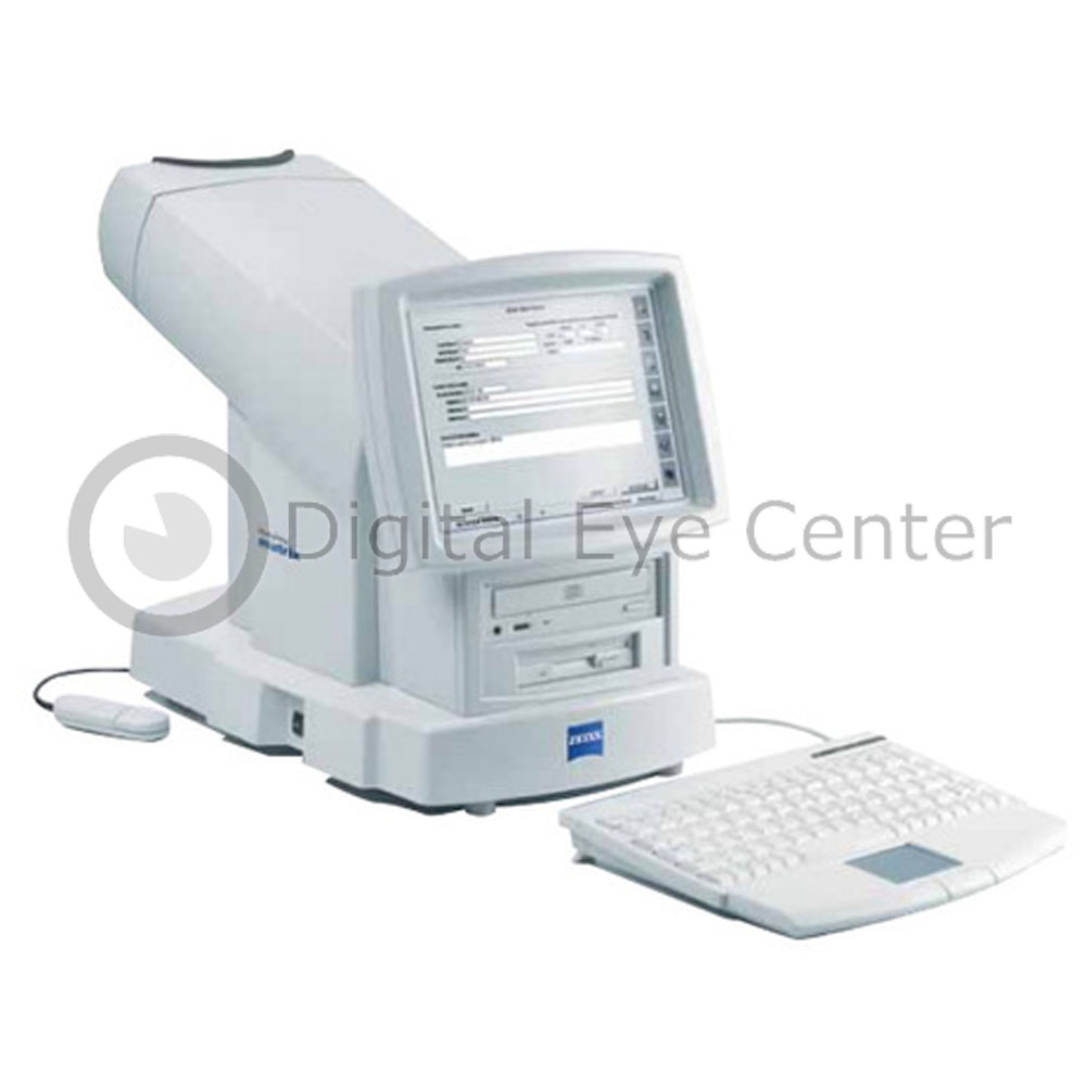
This perimeter utilizes Welch Allyn Frequency Doubling Technology.
Represents a significant breakthrough in Visual Field testing by combining early glaucoma detection capabilities, tools to characterize glaucoma, and basic glaucoma management tools.
The Humphrey Matrix also doubles as an in-office glaucoma screener with 35-second supra-threshold testing. Also provides up to 69 stimuli to characterize visual field defects to facilitate accurate diagnoses.
Comes equipped with Glaucoma Asymmetry Test and serial field overview software for comprehensive threshold exams and charting change over time, clinically validated by numerous studies.
Patient and practice friendly, the Zeiss Humphrey Matrix have a video eye monitoring feature that simplifies patient alignment and fixation control.
Additionally, when using the Matrix, an eye patch isn’t required, and trial lenses are only needed beyond +/- 3 diopters. It also comes with enhanced optics for 30° FOV.
The Zeiss Humphrey Matrix performs dependably in ambient light so there’s no need to darken the room. Humphrey Matrix test results are presented on an LCD color display and can be printed on an external color printer. Can store up to a million exams for historical analysis.
Only minimal training is required, and any staff member can operate the Matrix Perimeter. Compact, lightweight, and with only a small footprint, the Humphrey Matrix can be located almost anywhere in your practice.
alyzer Features:
• Excellent for characterizing Visual Field status and providing a basic glaucoma management platform
• Threshold and screening tests for comprehensive care
• Patient management software including Glaucoma Asymmetry Test and serial field overview for comprehensive exam results
• Clinical validation from numerous studies documenting the Humphrey Matrix’s diagnostic performance
• Easy and intuitive operation for users of any ability
Table Top and Unit arm included. If you need an electric table mount, let us know.
Power Cable
Instruction Manual
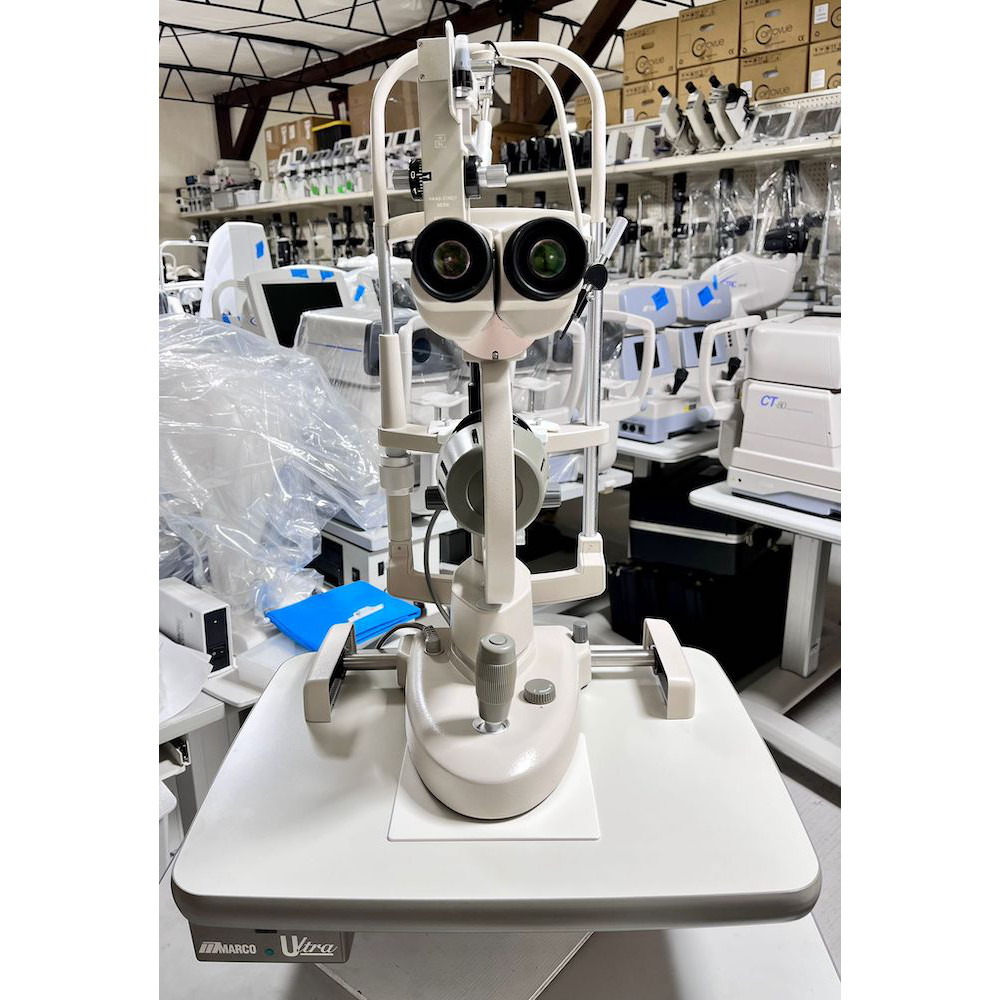
The Marco G2 slit lamp is an essential tool for any ophthalmologist or optometrist, as it provides a detailed examination of the anterior segment of the eye. With its advanced features and high-quality optics, the Marco G2 slit lamp is an excellent choice for any eye care professional looking to examine their patients’ eyes comprehensively.
Here are some of the key benefits of purchasing a used Marco G2 slit lamp from our eye center:
Affordable pricing: We offer used Marco G2 slit lamps at a significantly lower cost than purchasing new equipment. This means you can save money without sacrificing quality.
High-quality refurbishment: Our team of experts carefully inspects and refurbishes each piece of equipment to meet the manufacturer’s specifications. This ensures that you are getting equipment that is reliable, precise, and in excellent condition.
Wide range of used ophthalmic equipment: In addition to the Marco G2 slit lamp, we offer a variety of other used ophthalmic equipment, including phoropters, tonometers, and more. This means you can find everything you need in one convenient location.
Look no further than our eye center if you are in the market for a used Marco G2 slit lamp. Our equipment is fully refurbished to meet the manufacturer’s specifications, and our team of experts is dedicated to providing only the highest quality products and services. With our affordable pricing and wide range of used ophthalmic equipment, we are confident that you will find everything you need to provide the best possible care for your patients.
Table Top and Unit arm mount included. If you need an electric table mount, let us know.
Power Cable
Instruction Manual
The Marco G5 slit lamp is an essential tool for any ophthalmologist or optometrist, as it provides a detailed examination of the anterior segment of the eye. With its advanced features and high-quality optics, the Marco G2 slit lamp is an excellent choice for any eye care professional looking to examine their patients’ eyes comprehensively.
Here are some of the key benefits of purchasing a used Marco G5 slit lamp from our eye center:
Affordable pricing: We offer used Marco G5 slit lamps at a significantly lower cost than purchasing new equipment. This means you can save money without sacrificing quality.
High-quality refurbishment: Our team of experts carefully inspects and refurbishes each piece of equipment to meet the manufacturer’s specifications. This ensures that you are getting equipment that is reliable, precise, and in excellent condition.
Wide range of used ophthalmic equipment: In addition to the Marco G5 slit lamp, we offer a variety of other used ophthalmic equipment, including phoropters, tonometers, and more. This means you can find everything you need in one convenient location.
Look no further than our eye center if you are in the market for a used Marco G5 slit lamp. Our equipment is fully refurbished to meet the manufacturer’s specifications, and our team of experts is dedicated to providing only the highest quality products and services. With our affordable pricing and wide range of used ophthalmic equipment, we are confident that you will find everything you need to provide the best possible care for your patients.
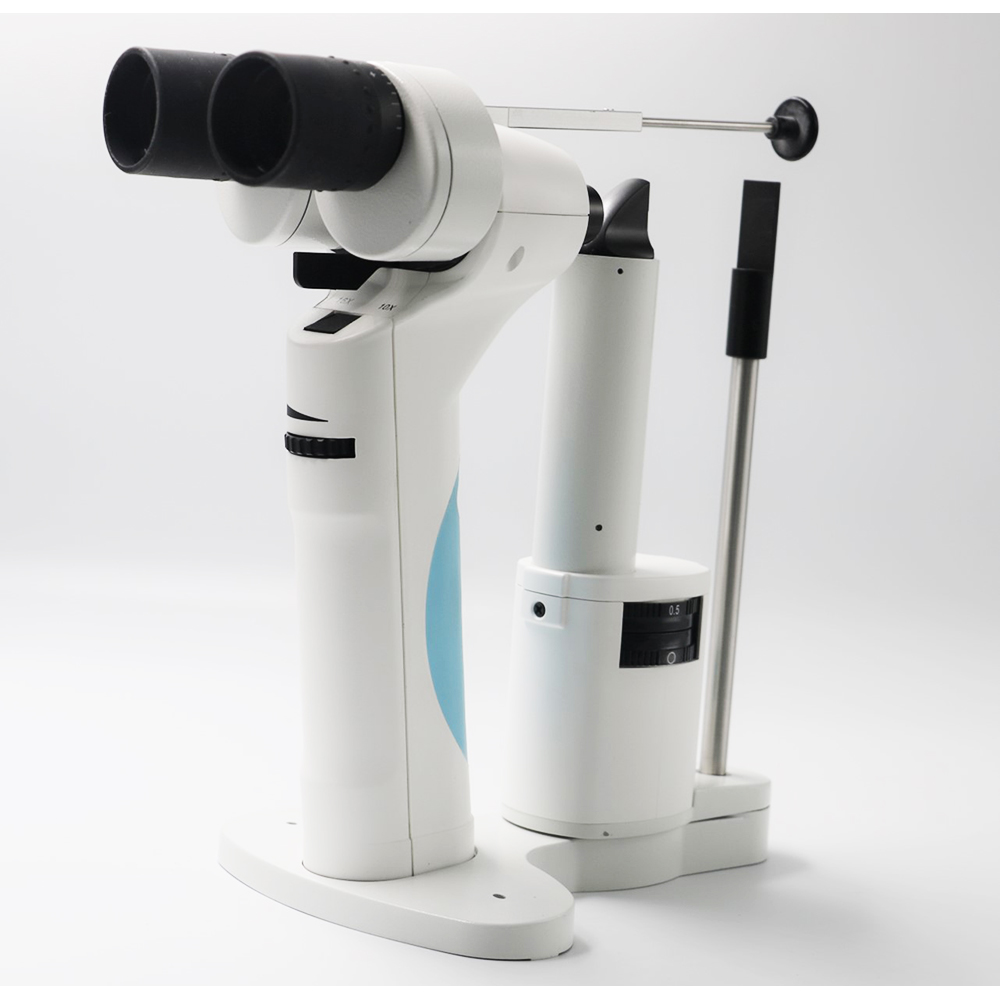
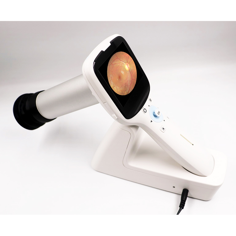
Its light weight make this a perfect equipment for the doctor that needs to take remote exams, or doesn’t have a desktop fundus camera.
It will integrate with our own iPad App for easy wireless transfer of pictures and patients information.
It features a 4-hour battery and sd card slot, were you can save up to 80,000 HD images.
The Digital Eye Portable Fundus Camera is perfect for fundus imaging, diagnosis and disease screening.
This is a hand held retinal camera for fundus imaging, diagnosis and especially for fundus disease screening, and easy to obtain high definition fundus image. Applied to rapid screening, out diagnosis, bedside diagnosis and remote medical treatment.
Characteristics
Non-Mydriatic
45 degrees viewing angle
WIFI Transmission to PC and iPad.
Applications of Basic Fundus Screening
Small Clinics
Mobile and remote screening programs
Disabled and elderly patients
Fast screening for children
ROP test for premature infants under 2.5 kg
Real-time monitoring for cerebral edema after brain surgery
Monitoring all stages of diabetic retinopathy
Convertible to desktop with slit lamp adapter
Patient information management software
Applications outside Ophthalmology
Retinal Photography as a wider field of practice beyond Ophthalmology. Here is a brief list of other fields:
Internal Medicine
Retinal micro-vascular abnormalities and cardiovascular disease
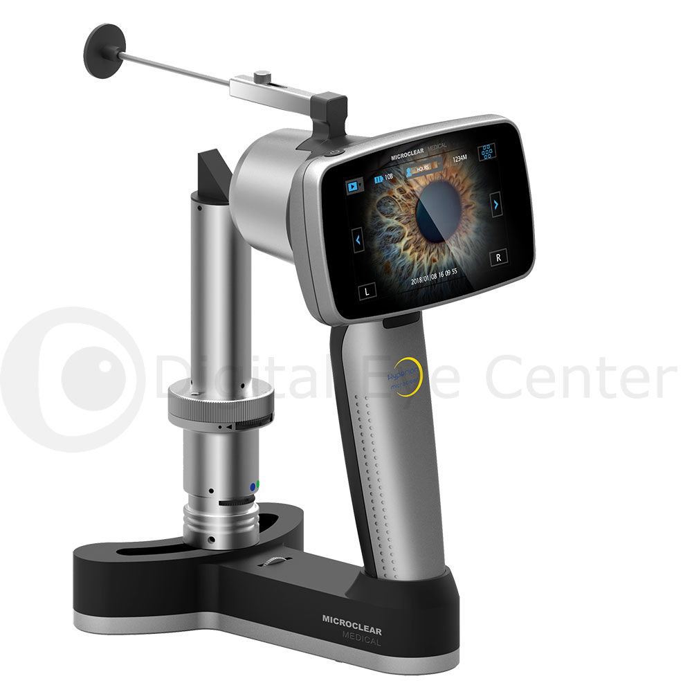
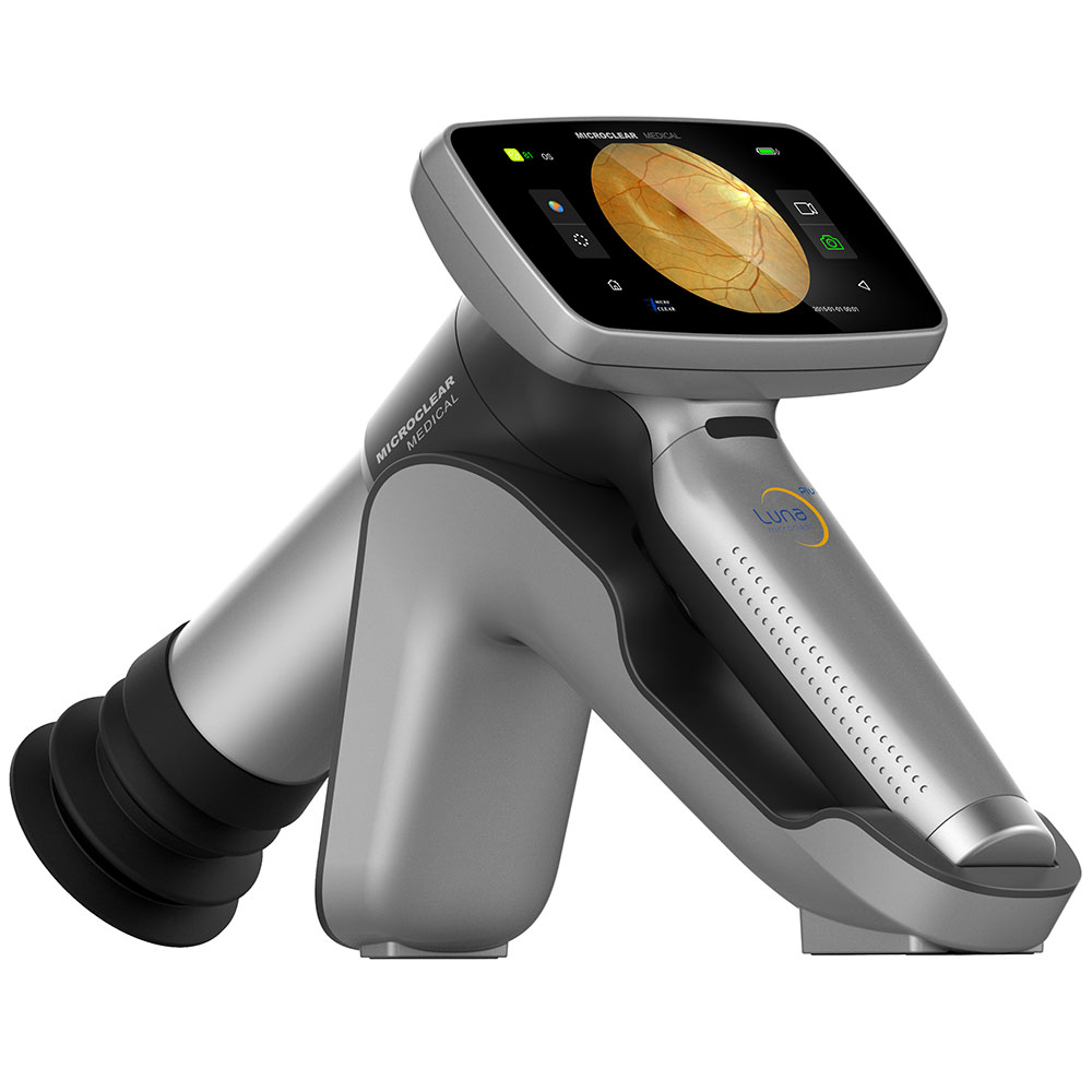
Its light weight make this a perfect equipment for the doctor that needs to take remote exams, or doesn’t have a desktop fundus camera.
It features a 6-hour battery and 16gb of internal memory , were you can save up to 3,300 HD images.
The Microclear Luna Portable Fundus Camera is perfect for fundus imaging, diagnosis and disease screening.
This is a hand held retinal camera for fundus imaging, diagnosis and especially for fundus disease screening, and easy to obtain high definition fundus image. Applied to rapid screening, out diagnosis, bedside diagnosis and remote medical treatment.
Features of the Portable Fundus Camera
Non-Mydriatic and Minimum pupil size 2.5 mm
Low light LED flash and 480×800 HD touch screen
Autofocus and 8 target fixation
16 mpx JPG images
Android System for easier operation and data storage
Wireless Transfer
DICOM interface
Weight only 560 g
Wireless Printing
Applications outside Ophthalmology
Retinal Photography as a wider field of practice beyond Ophthalmology. Here is a brief list of other fields:
Internal Medicine
Retinal micro-vascular abnormalities and cardiovascular disease
Filters: Red free, cobalt blue, Red free, cobalt blue, heat absorbing, neutral density
Movement Ranges: Longitudinal: (in/out) 90 mm – Lateral: (left/right) 107 mm 100 mm – Vertical: (up/down) 30 mm 30 mm
Chin Rest Range 80 mm 80 mm
VAC 110/220 VAC.More Resources
Features
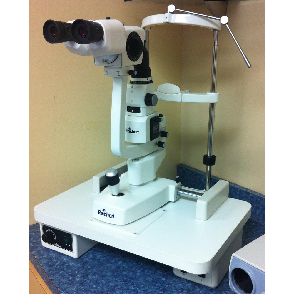
One of the key features of the Reichert Xcel 200 is its advanced optical system, which includes a Galilean converging microscope with a 3-step magnification changer. This allows for clear, high-resolution images at a range of magnifications, from 10x to 25x.
The instrument also features a 14mm slit aperture with continuously adjustable length and width and a 12-degree converging angle for improved depth perception. This makes it ideal for a variety of diagnostic procedures, including anterior segment examinations, contact lens fittings, and glaucoma evaluations.
In addition, the Reichert Xcel 200 has a built-in yellow filter for enhanced visualization of subtle details and a cobalt blue filter for assessing corneal abrasions and foreign bodies.
Benefits
Adding the Reichert Xcel 200 Slit Lamp to your practice can provide more accurate diagnoses and treatments for a range of eye conditions. Its high-quality optics and advanced features allow for detailed examination of the anterior and posterior segments of the eye, while its comfortable design ensures a positive patient experience.
Furthermore, choosing a used instrument like the Reichert Xcel 200 can be a cost-effective way to expand your practice without breaking the bank. At Digital Eye Center, we carefully inspect and refurbish all of our used equipment to ensure that it meets our high standards for quality and reliability.
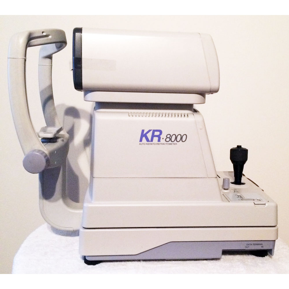
The innovative design of this used Topcon KR-8000 enables accurate, reliable refraction and keratometric measurements within a pupil dilation as narrow as 2.0mm. This means easier and more precise diagnostic results when dealing with glaucoma, asymmetric pupils, or elderly patients.
Totally reliable and totally accurate with Topcon’s Rotary Prism Measuring System, our advanced rotary prism principle is able to obtain highly reliable data. The off-centered ring target is able to measure areas previously occluded by a small pupil. The prism angle enables a much wider fundus area to be measured. Accordingly, the measurement image provides extremely reliable data.
Objective Refractometer Mode Sphere -25 to +22D in 0.25D step (0.12D step available)
Cylinder 0 to ±8D in step 0.25D (0.12D step available)
Axis 1° to 180° in 1° and 5° step
Minimal pupil diameter 2.0mm ø
Method of relaxation Automatic fogging Charts for object testing
Table Top and Unit arm included. If you need an electric table mount, let us know.
Used Haag Streit 870 Tonometer
Power Cable
Instruction Manual
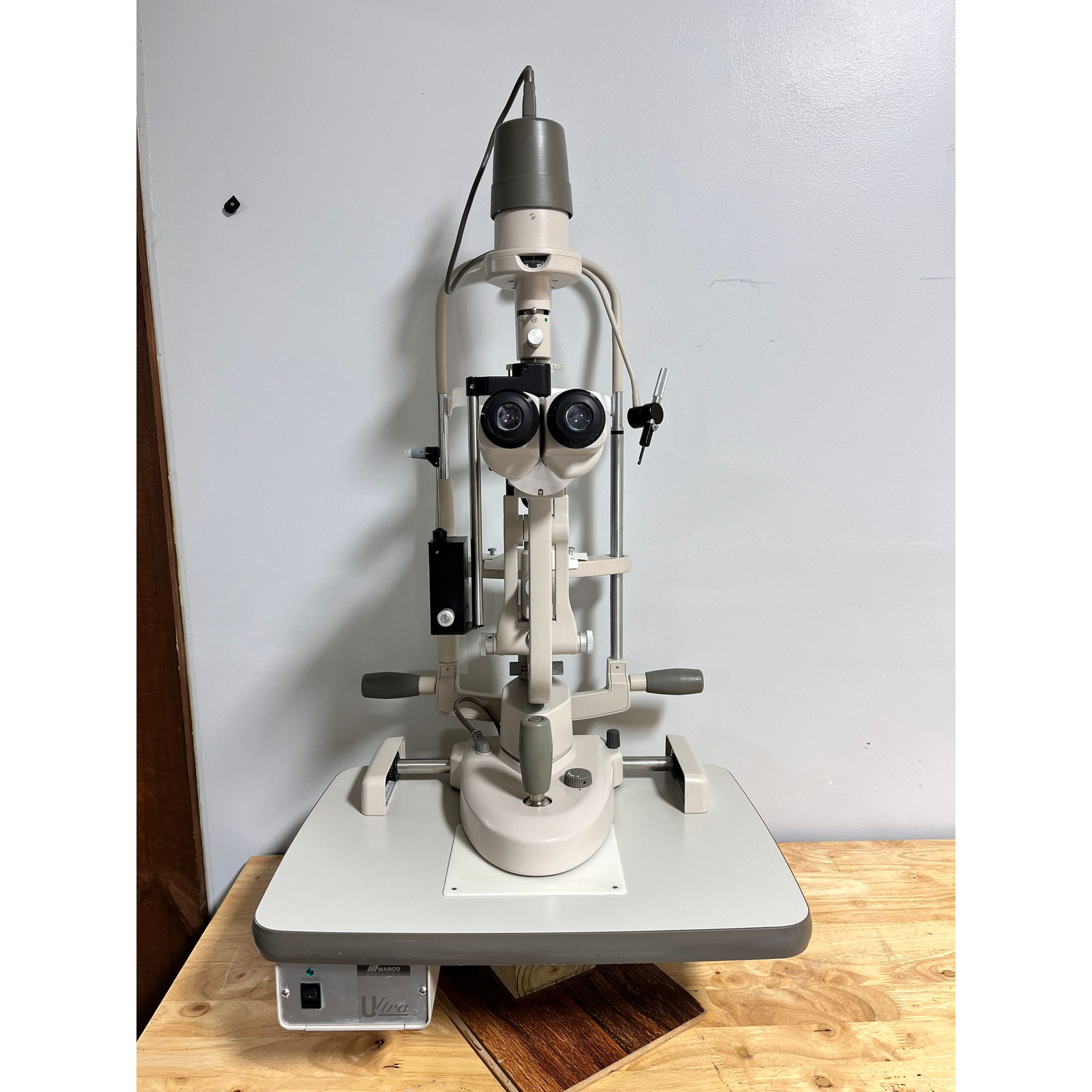
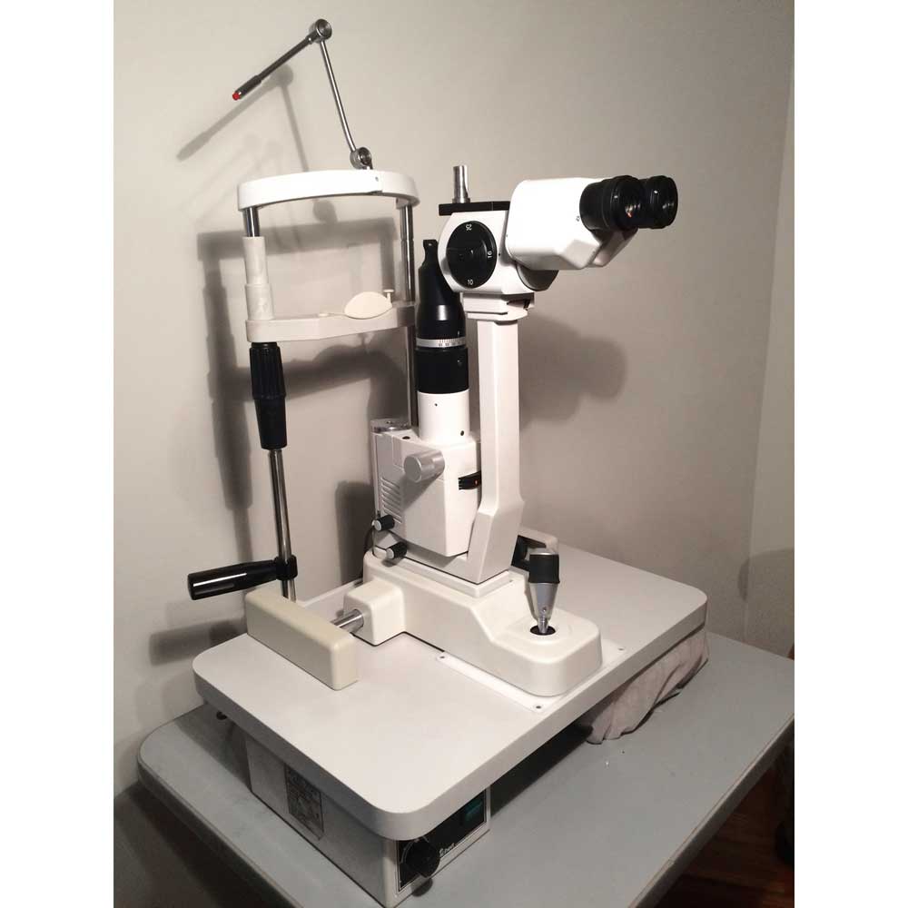
Used Topcon SL 2G Slit Lamp Specifications
The Topcon SL-2G slit lamp uses an LED bulb instead of a traditional one inside a compact illumination unit. LED technology results in uniform and consistent illumination and is more eco-friendly because LED lasts 100x longer than conventional halogen bulbs.
This ophthalmic slit lamp features three magnifications (10x, 16x, and 25x) for a comprehensive examination. The adjustable slit mechanism has up to 14 mm of aperture, and the slit angle rotates to 180 degrees. A simple lever movement can insert red-free and cobalt-blue filters.
Features
LED illumination with bulbs that last 100x longer than traditional halogen bulbs
Affordable option with all necessary functions for a comprehensive eye exam
Bright, clear, and distortion-free view from Topcon’s specially treated lenses
Easy and comfortable to use for both the patient and operator because of the ergonomic design
Filters enhance observation with cobalt blue for fluorescein staining and IOP measurements and red-free for blood vessel and nevus observation.
Omni-directional joystick and elevation mechanism for smooth movement and use.
0 to 180 slit angle rotation.
How Does the Topcon SL-2G Slit Lamp Compare to Other Brands?
When it comes to slit lamps, there are many brands to choose from. However, the Topcon SL-2G slit lamp stands out from the crowd for several reasons. Here are a few key differences between the Topcon SL-2G slit lamp and other popular brands:
Illumination – The Topcon SL-2G slit lamp provides superior illumination compared to many other brands. This makes seeing every detail of the patient’s eye easier, even in low-light conditions.
Optics – The Topcon SL-2G slit lamp is equipped with high-quality optics that provide a clear and detailed eye view. Many other brands use lower quality optics that can compromise the accuracy of your diagnoses.
Ease of Use – The Topcon SL-2G slit lamp is designed with user-friendliness in mind. The joystick and aperture adjustments are intuitive and easy to use, making it quick and easy to get the perfect view of the eye.
Durability – The Topcon SL-2G slit lamp is built to last. It’s made from high-quality materials that can withstand the wear and tear of daily use in a busy eye care practice.
Versatility – The Topcon SL-2G slit lamp is versatile and can be used for various examinations. This includes anterior segment examinations, contact lens fittings, and more.
Overall, the Topcon SL-2G slit lamp is a top choice for eye care professionals who want the best possible equipment for their practice.