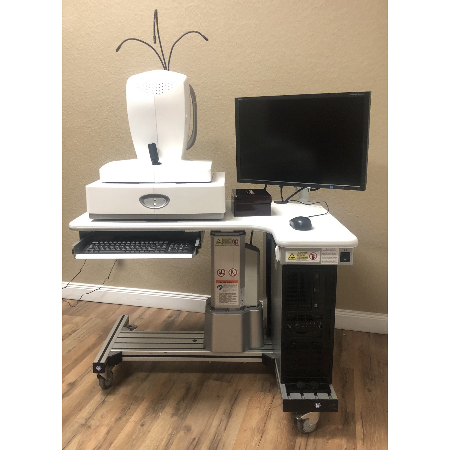
Specifications for Cirrus 4000
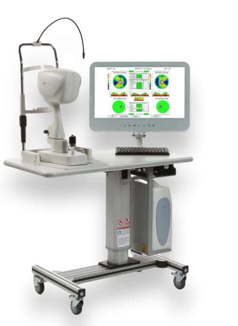
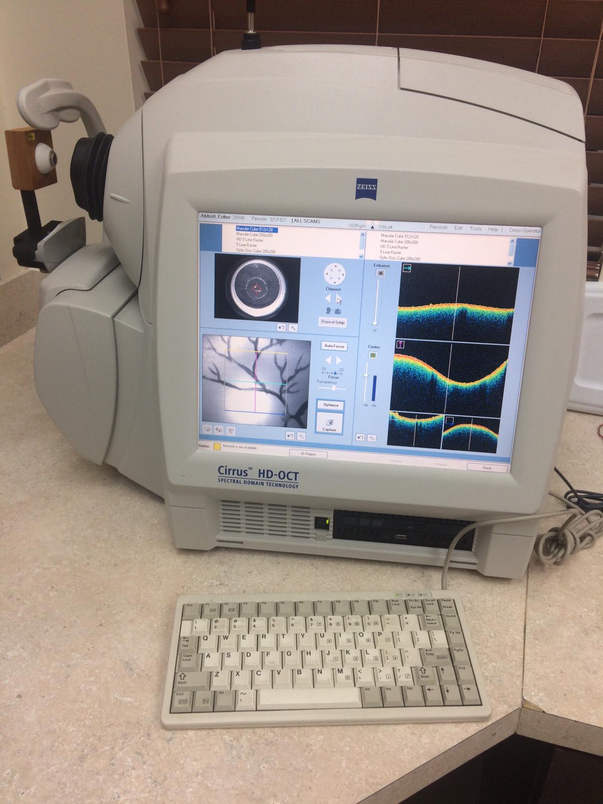
The ZEISS Cirrus HD-OCT 4000 (Cirrus HD-OCT or Cirrus) enables examination of the posterior and anterior of the eye at an extremely fine spatial scale, without surgical biopsy or even any contact with the eye.
The Cirrus HD-OCT builds on and refines the retinal imaging technology first introduced with the ZEISS Stratus OCT™.
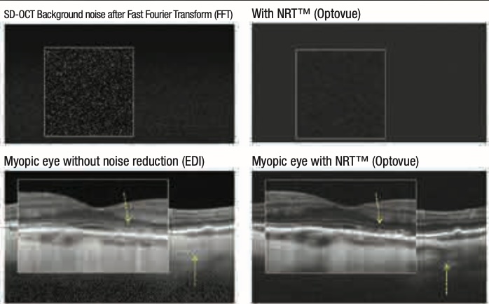
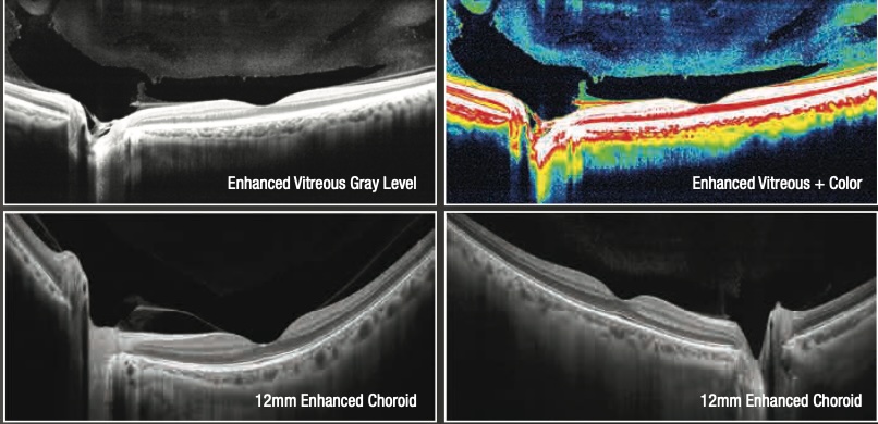
Employing the advanced imaging technology of spectral-domain optical coherence tomography, Cirrus HD-OCT acquires OCT data about 70 times faster (27,000 vs. 400 A-scans per second) and with better resolution (5 μm vs. ~10 μm axial resolution in tissue), compared to first-generation OCT technology.
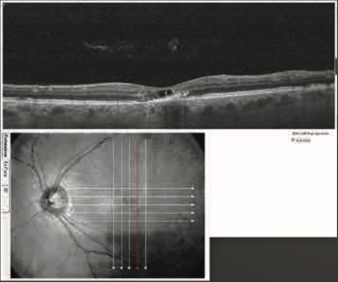
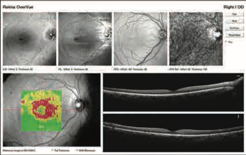
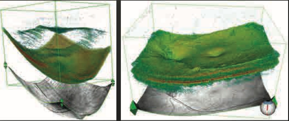
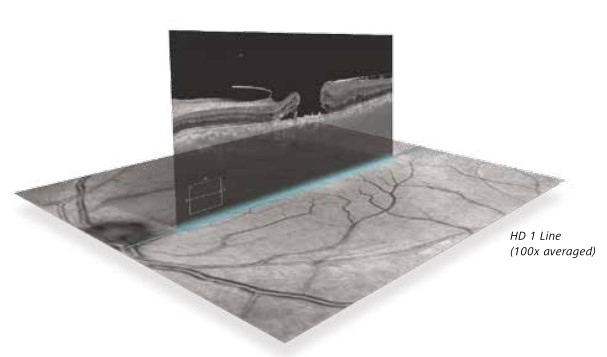
Cirrus acquires whole cubes of OCT image data, composed of hundreds of line scans, in about the same time as Stratus acquires a six-line scan. You can view these data cubes in three planes, or through three dimensions, giving you access to an extensive amount of retinal image data in one scan.
Specifications for Cirrus 4000
The Cirrus HD-OCT with Retinal Nerve Fiber Layer (RNFL), Macular, Optic Nerve Head, and Ganglion Cell Normative Databases is indicated for in-vivo viewing, axial cross-sectional, and three-dimensional imaging and measurement of anterior and posterior ocular structures.
Indications for Use
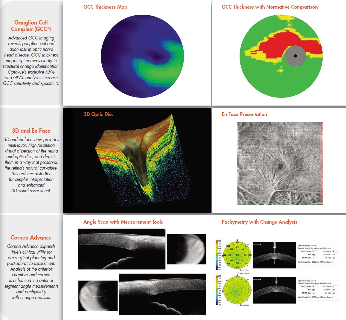
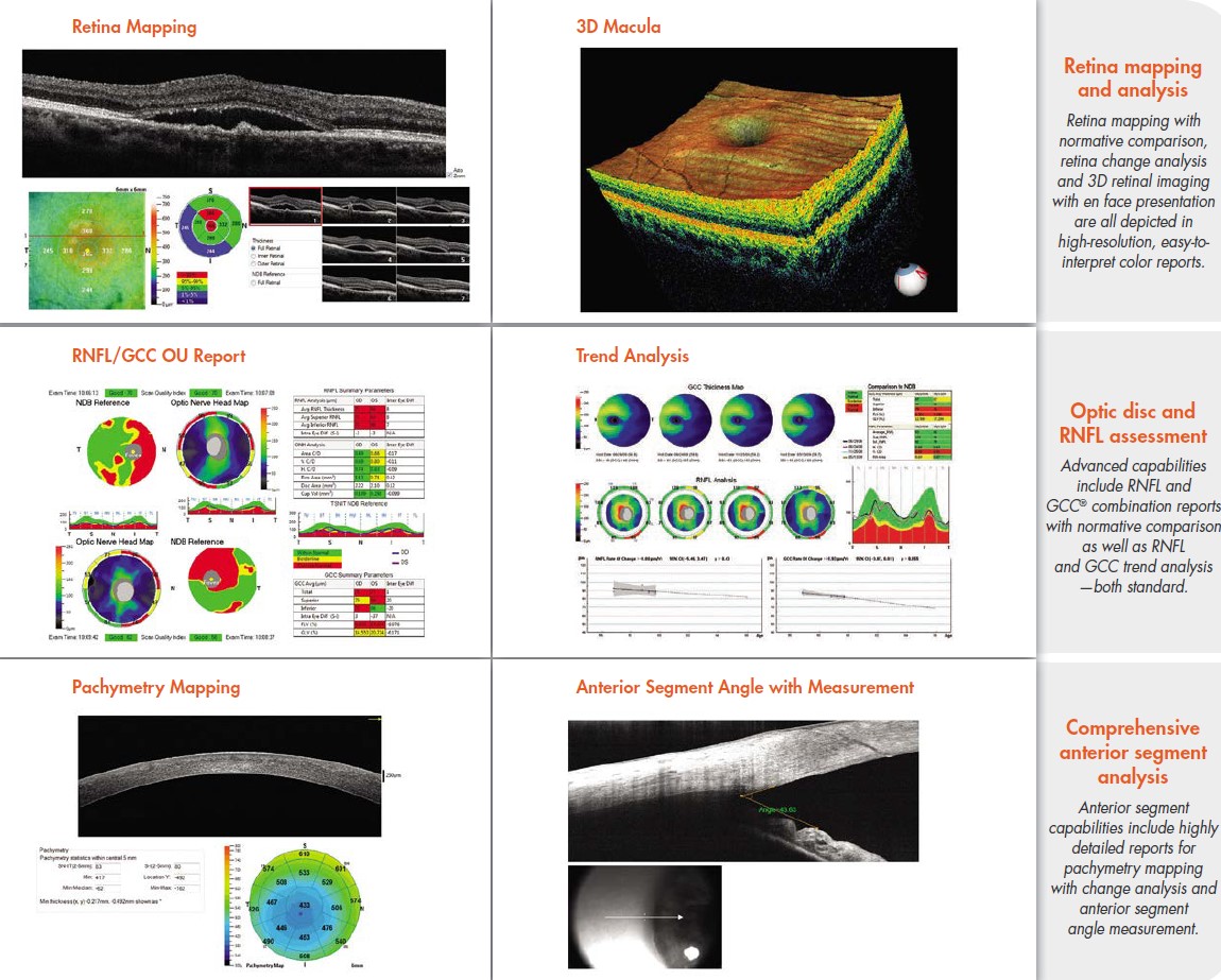
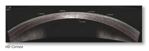
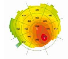
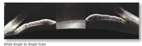
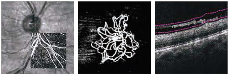
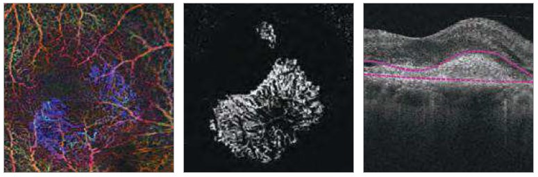
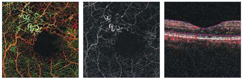
The Cirrus HD-OCT is a non-contact, high-resolution tomographic and biomicroscopic imaging device. It is indicated for in-vivo viewing, axial cross-sectional, and three-dimensional imaging and measurement of anterior and posterior ocular structures, including cornea, retina, retinal nerve fiber layer, ganglion cell plus inner plexiform layer, macula, and optic nerve head.
The Cirrus normative databases are quantitative tools for the comparison of retinal nerve fiber layer thickness, macular thickness, ganglion cell plus inner plexiform layer thickness, and optic nerve head measurements to a database of normal subjects.
The Cirrus HD-OCT is intended for use as a diagnostic device to aid in the detection and management of ocular diseases including, but not limited to, macular holes, cystoid macular edema, diabetic retinopathy, age-related macular degeneration, and glaucoma.
Note: The Cirrus HD-OCT is not intended to be used as the sole diagnostic for disease.
Essential Performance
The Essential Performance of the instrument is to provide accurate measurements of the anterior and posterior ocular structure.
Patient Population
The Cirrus HD-OCT may be used on all adults in need of diagnostic evaluation of the eye. This includes (but is not limited to) patients with the following disabilities or challenges:
• Wheelchair user
• Very low or not measurable visual acuity
• Fixation problems
• Postural problems
• Deafness
• Large body, but not those above 99th percentile based on anthropomorphic data
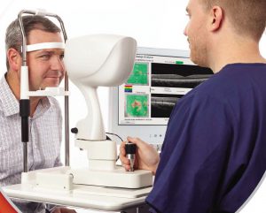
There is a general requirement that the patient is able to sit upright and be able to place their face in the chin and forehead rest of the instrument (with or without supplemental human or mechanical support).
Cirrus HD-OCT is designed for in-vivo viewing, axial cross-sectional, and three-dimensional imaging and measurement of anterior and posterior ocular structures.