$37,000.00
Lease from $892/month. Apply HERE
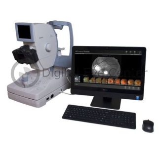
This Cirrus 5000 Quad Core is refurbished equipment with a six-month warranty. It includes the anterior segment, Windows 10, and Table.
Includes:
- Cirrus 5000 OCT with Angioplex
- Quad Core Processor
- Windows 10
- Table
- six months warranty
- Optional: Premier Lens: Add $4,000
Only 1 left in stock (can be backordered)
Description
The Zeiss Cirrus 5000 OCT improves on the Cirrus 4000 model in hardware and software. It retains the essential features of the 4000 model and upgrades on hardware and Smart HD Scans.
Eye motion artifacts are reduced with FastTrac retinal tracking, which enables tracking of the current scan to the position of a previous scan, which, as a result, provides a better progression analysis.
The monitor has been improved, too, with a 19-inch display for high-definition scans.
AngioPlex OCT Angiography
For the first time, AngioPlex allows visualizing vascular and structural information from a single non-invasive scan. One that makes eye care’s leading clinical OCT platform an unprecedented tool for acquiring ultra-clear color-depth-resolved 3D microvascular retina imaging.
AngioPlex technology revolutionizes clinical practice by making the visualization of the microvasculature of the retina a routine part of everyday care.
- New vascular information
- Ultra-clear 3D microvascular visualizations powered by OMAG
- OMAG detects the motion of red blood cells within sequential OCT B-scans repeatedly performed at the same location
- Depth of retinal vasculature color-coded for ease of visual assessment
- Enhanced workflow
- Ideal non-invasive, dye-free angiography
- Single-scan simplicity: capture OCT angiography with just one scan
- Real-time tracking with enhanced FastTrac ensures artifact-free scans and precise location identification during follow-up visits.
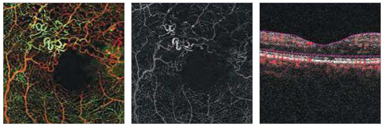
| Diabetic Retinopathy (DR) – AntioPlex(TM) maps visually isolate the neovascularization elsewhere (NVE) located in the vitro-retinal interface (VRI) |
|
|
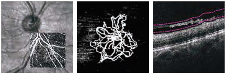
| Age-Related Macular Degeneration (AMD) – AngioPlex maps reveal choroidal neovascularization( CNV) in AMD. |
|
|
|
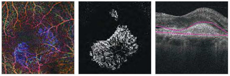
Branch Retinal Vein Occlusion (BRVO) – AngioPlex maps visualize vascular abnormalities and areas of non-perfusion due to vein occlusions
|
 |


NEW PanoMap Analysis
Wide-field structural damage assessment for glaucoma
-
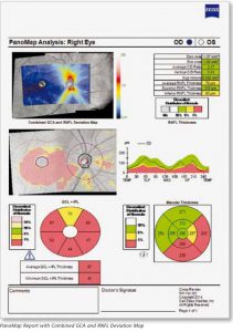
Zeiss PanoMap The new PanoMap wide field analysis displays structural data for the entire posterior pole. RNFL, ONH, and GCO metrics show the extent of structural damage.
- At-a-glance insight — A single analysis for integrated insights into early pathologies
- Backward-compatible –PanoMap uses existing Macular Cube and Optic Disc Cube scans to provide a wide-field view of the posterior pole without altering scan protocols.

NEW Anterior Segment Premier Module from Zeiss
The first retinal OCT with complete anterior chamber imaging and measurements
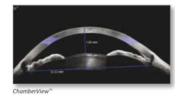

ChamberView image – Chamberview provides an expansive 15.5 mm wide view of the anterior chamber with objective tools for measuring anterior segment ocular structures.
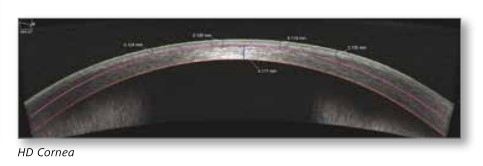

HD Cornea Scan—This is a 9 mm high-resolution scan that includes versatile tools for measuring residual stromal bed thickness, LASIK flap thickness, and other corneal structures.
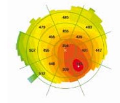

Pachymetry Map –9 mm pachymetry map highlights corneal irregularities and identifies the thinnest point for refractive surgery screening.
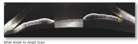
NEW OCT Goniometry
A non-contact method to help identify patients at risk of angle closure glaucoma

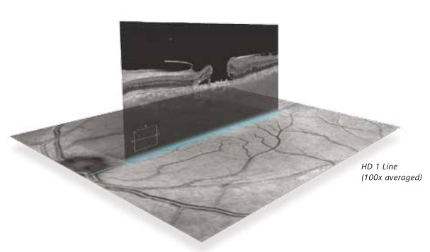
NEW Smart HD Scan Patterns
Targeted visualizations of critical anatomy.
The automatic centering of scans ensures that the fovea is seen in each patient.

Technical Specifications
| OCT Imaging | |
| Methodology | Spectral Domain OCT |
| Optical source | Superluminescent diode (SLD), 840 nm |
| Scan speed | 27,000 – 68,000 A-scans per second |
| A-scan depth | 2.0 mm (in tissue), 1024 points |
| Axial resolution | 5 µm (in tissue) |
| Transverse resolution | 15 µm (in tissue) |
| Fundus Imaging | |
| Methodology | Line scanning ophthalmoscope (LSO) |
| Live fundus image | During alignment and OCT scan |
| Optical source | Superluminescent diode (SLD), 750 nm |
| Field of view | 36 degrees W x 30 degrees H |
| Frame rate | > 20 Hz |
| Transverse resolution | 25 µm (in tissue) |
| Iris Imaging | |
| Methodology | CCD camera |
| Resolution | 1280 x 1024 |
| Live iris image | During alignment |
| Electrical and Physical | |
| Weight | 80 lbs (36 kg) |
| Dimensions of instrument | 26L x 18W x 21H (in) 65L x 46W x 53H (cm) |
| Dimensions of table | 39L x 22W (in) 99L x 56W (cm) |
| Fixation | Internal, external |
| Internal fixation focus adjustment | -20D to +20D (diopters) |
| Electrical rating (115V) | Single Phase, 100–120V~ systems: 50/60Hz, 5A |
| Electrical rating (230V) | Single Phase, 220–240V~ systems: 50/60Hz, 2.5A |
| Internal Computer | |
| Operating system/processor | Windows ® 7, 4 th generation i7 Intel ® processor |
| Memory | 16 GB |
| Hard drive/internal storage | ≥ 2 T > 200,000 scans |
| Display | Integrated 19“ color flat panel display |
| USB ports | Six ports |
Additional information
| Brand |
|---|