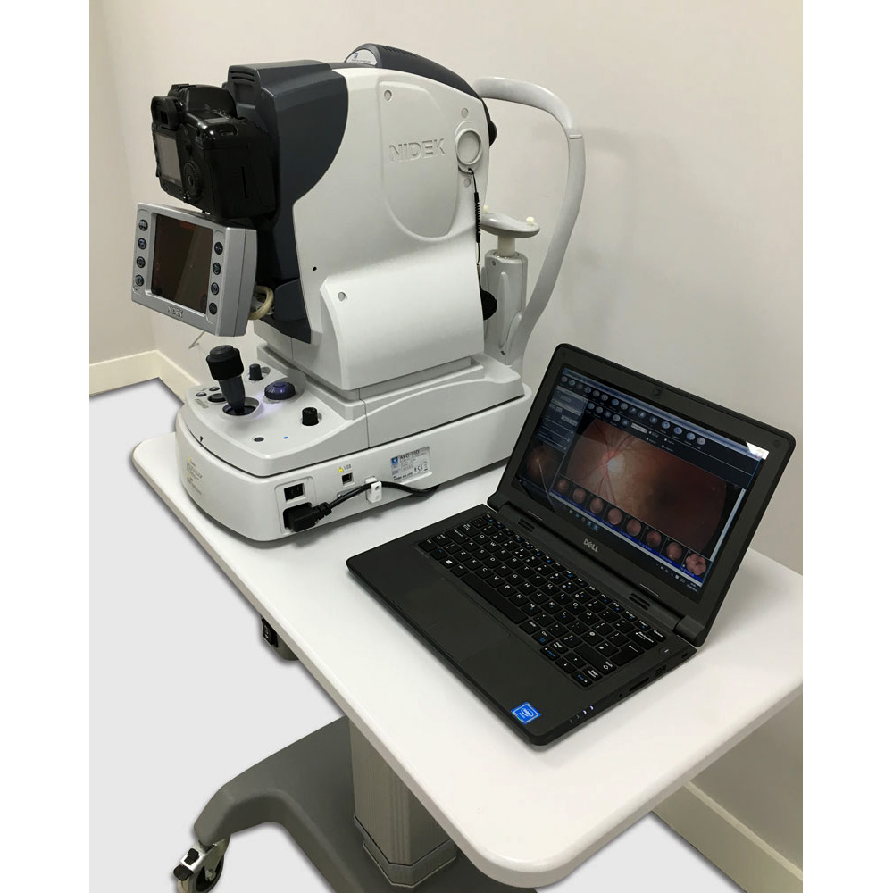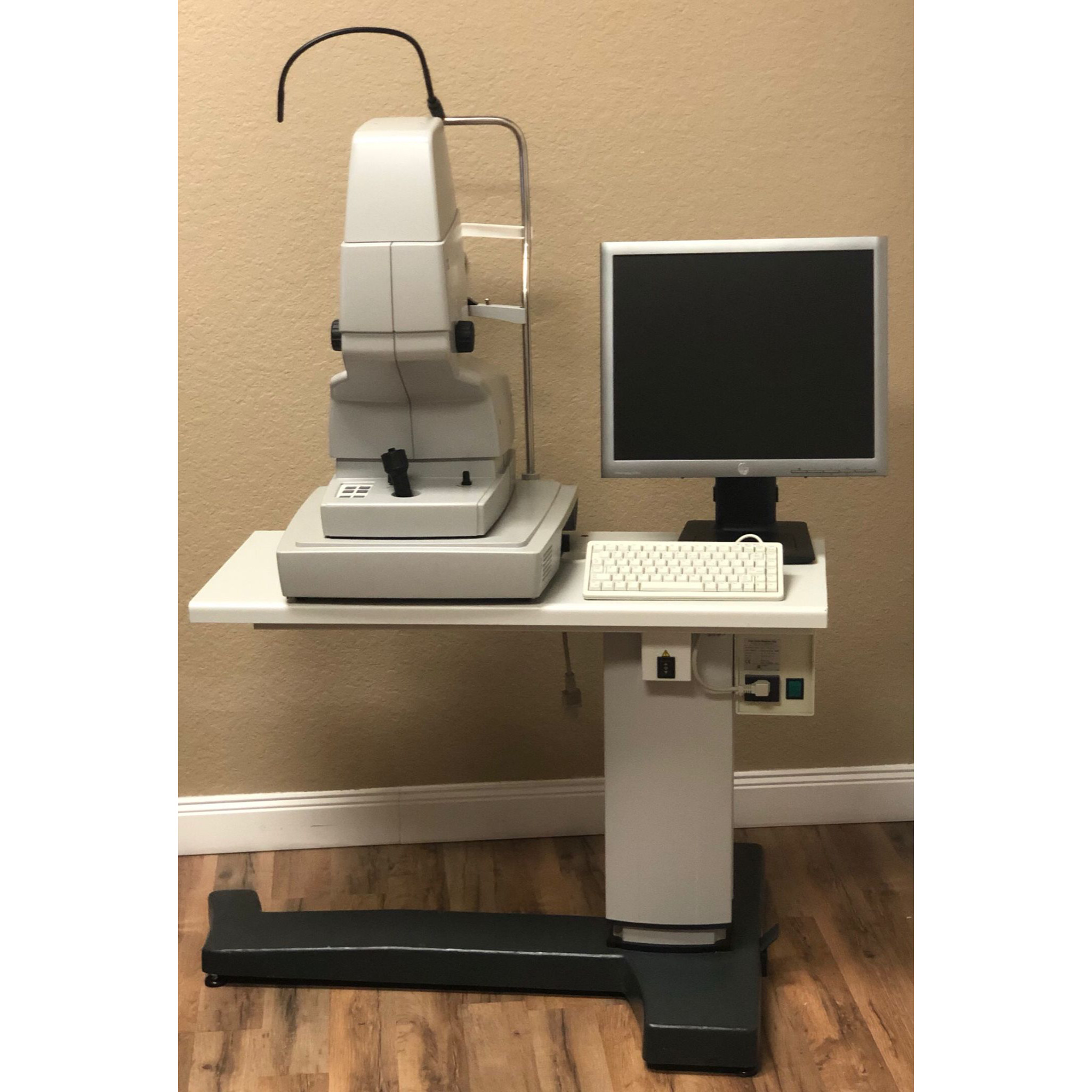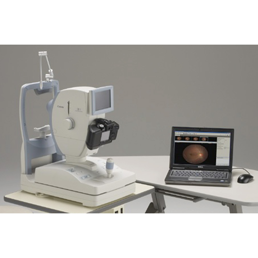
This Refurbished Canon CR-1 NM Fundus Camera System includes:
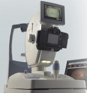
- Canon CR-1 NM Fundus Camera
- Digital Camera Canon DSLR
- Retinavue Software
- Laptop with Windows 10
- 6 months warranty
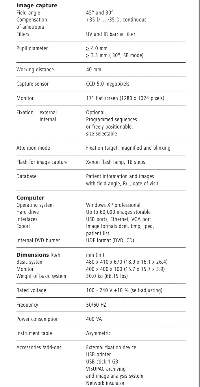
Specifications of the Canon CR-1 NM Fundus Camera
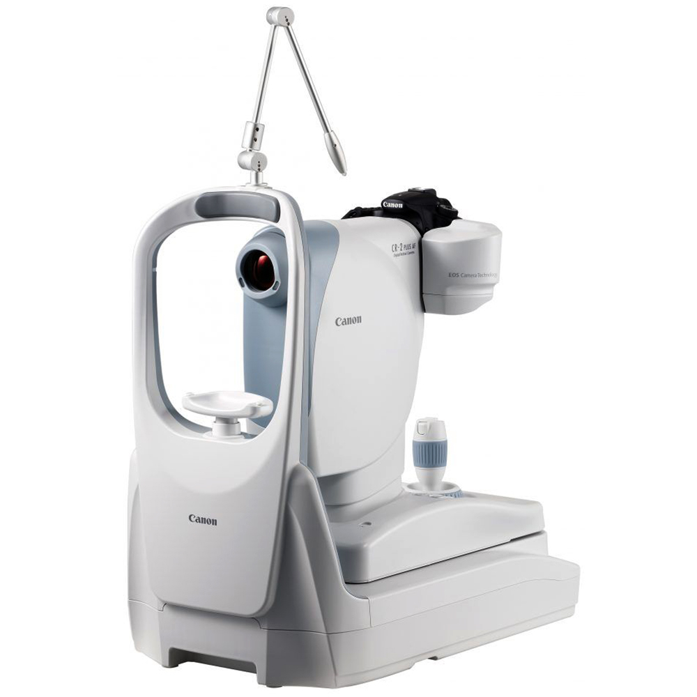
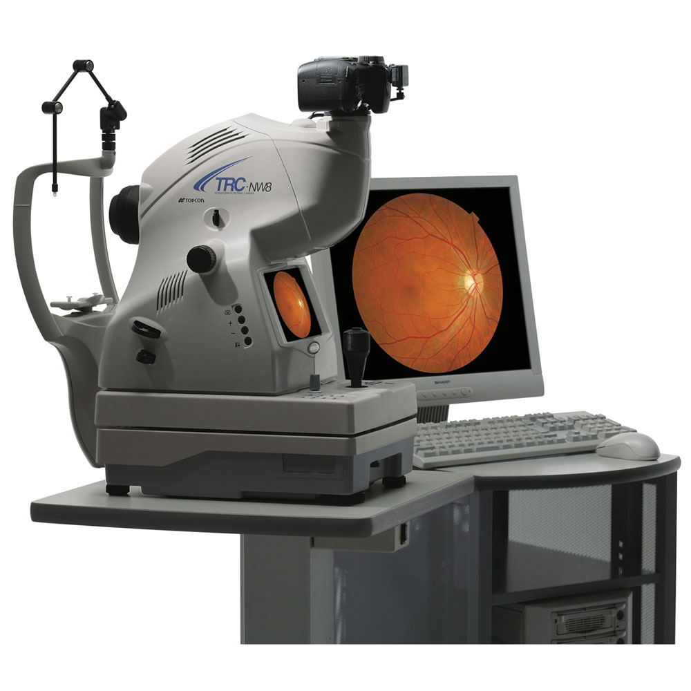
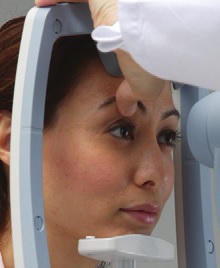
The State-of-the-Art in Non-Mydriatic Retinal Imaging, Ergonomically Designed.
The Non-Myd CR-1 features high-performance specs in an ergonomic, easy-to-use design to provide enhanced quality, efficiency and comfort during retinal exams.
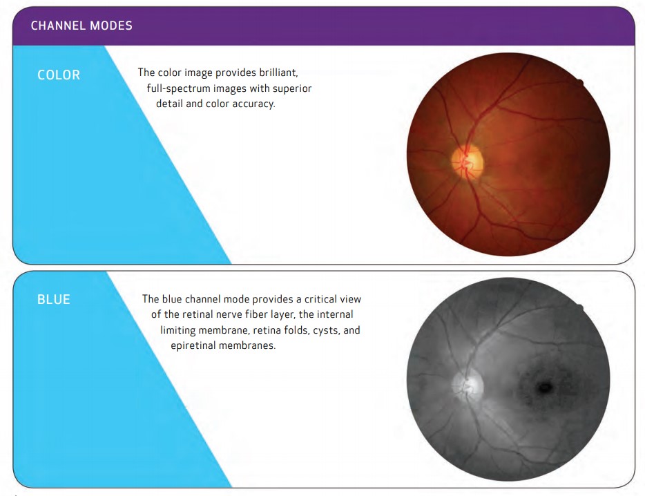
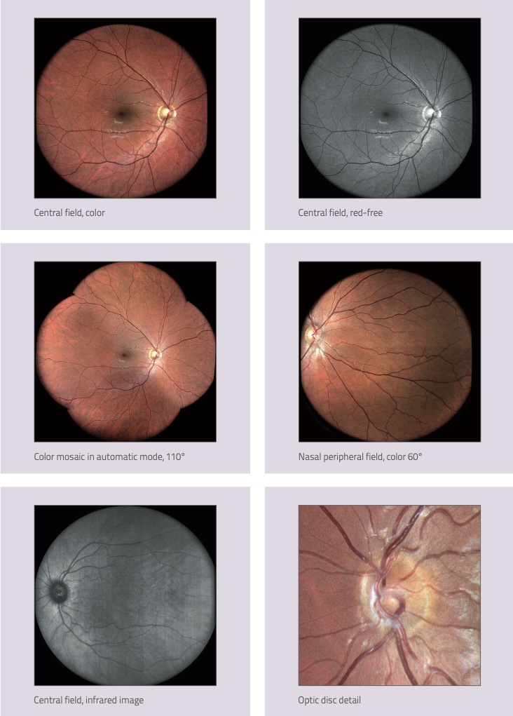
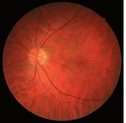
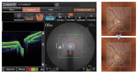
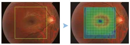
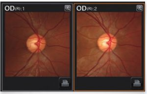
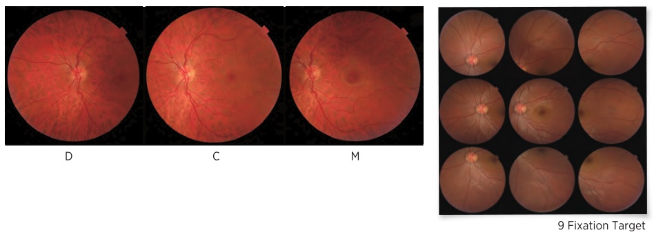
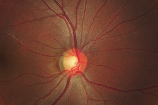
High-quality, high-resolution images
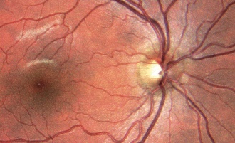
The CR-1 features a redesigned optical system that achieves extremely detailed, high-resolution diagnostic images of the retina for accurate detection and monitoring of ocular conditions including diabetic retinopathy, glaucoma, and macular degeneration.
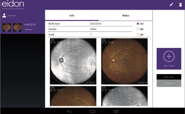
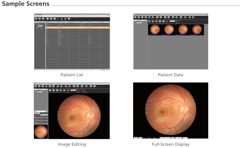
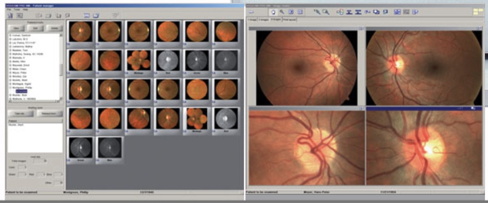
The high pixel count of the attached EOS digital SLR camera delivers detailed image quality even when magnified. Once captured, images are transferred to a connected PC for review.
Comfortable, ergonomic design

The all-new design of the CR-1 integrates advanced specifications into an ergonomic unit that facilitates operation by motorizing procedures usually performed by hand.
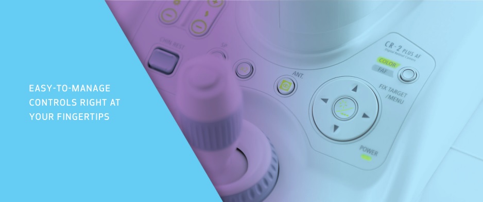
The intuitive controls, viewing monitor, and the streamlined form have all been designed for improved ease of use and comfort. It’s also patient-friendly. Because retinal images are easy to obtain, exams can be completed in less time.
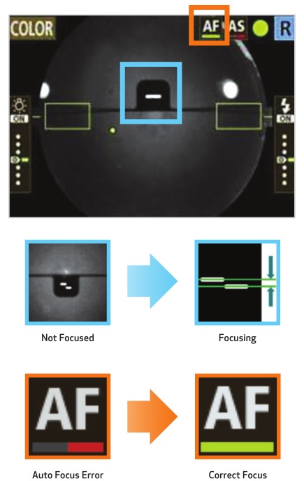
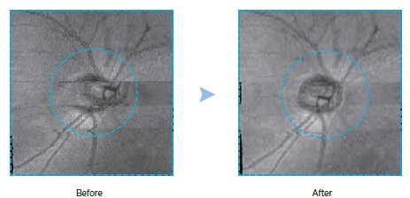
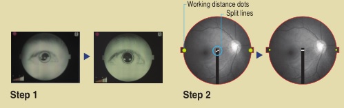
Easy alignment and focusing
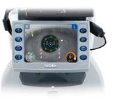
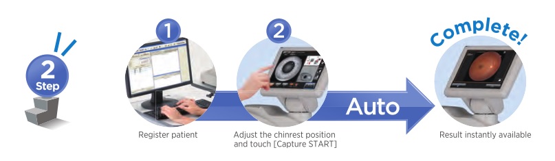
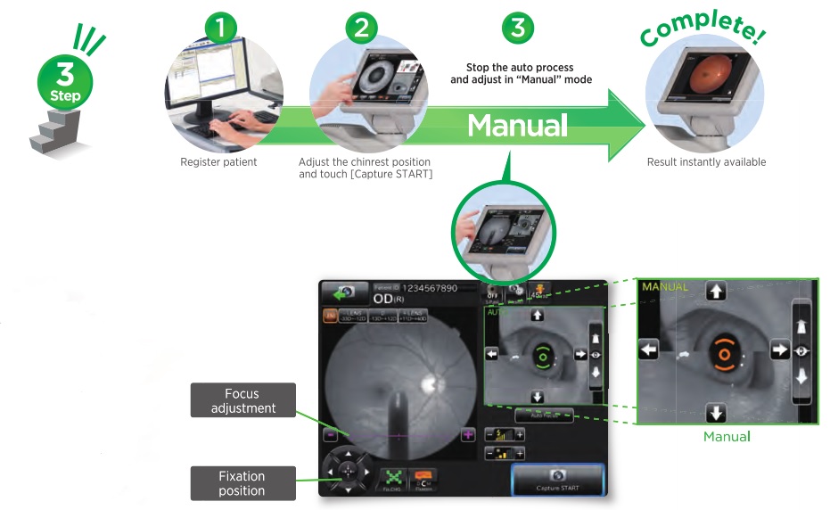
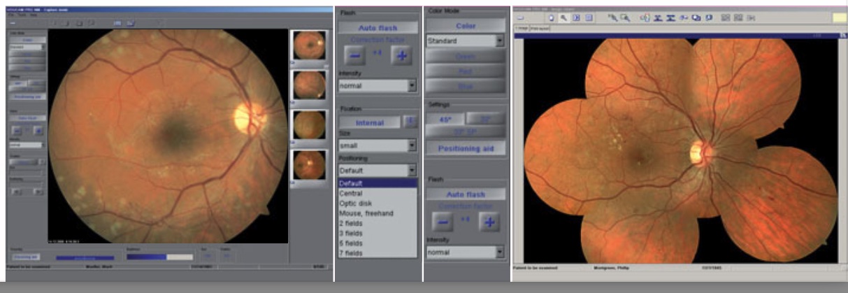
Image capture is fast and easy thanks to a simple two-step procedure. First, you align the two halves of a split pupil image. Then you switch to the retinal display, and adjust the split lines and working distance dots to achieve the correct focus and distance to the retina for clear, sharp images every time.

True 45-degree image

2x digital magnification
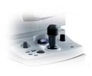
Motorized chin rest

The motorized chin rest can be moved up and down to accommodate the patient’s height using a pair of buttons located on the operation panel.
Front patient protection cover
The smooth front cover panel protects the patient from the instrument’s moving parts.
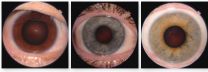
Small Pupil Mode
Photograph through undilated pupils as small as 3.7 mm.
Shorter working distance
Flash intensity adjustment, plus Low Flash Mode
Nine steps of flash adjustment are available, in addition to a low flash mode. Low flash intensity makes it easy to retake photos or take images of both eyes, when necessary.
Internal fixation target adjustment
The finely calibrated internal eye fixation target of the CR-1 provides the patient with a fixed, consistent point of focus throughout the image capture procedure, making it quick and easy to achieve a clear and stable image. The fixation target LED can be freely adjusted via the operation panel to position the eye exactly as desired. One push of a button returns the target to its default position.