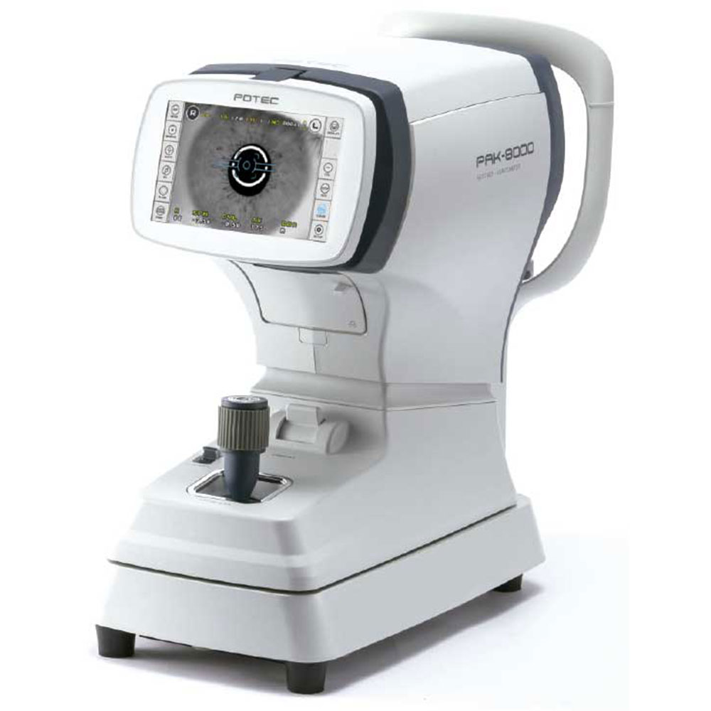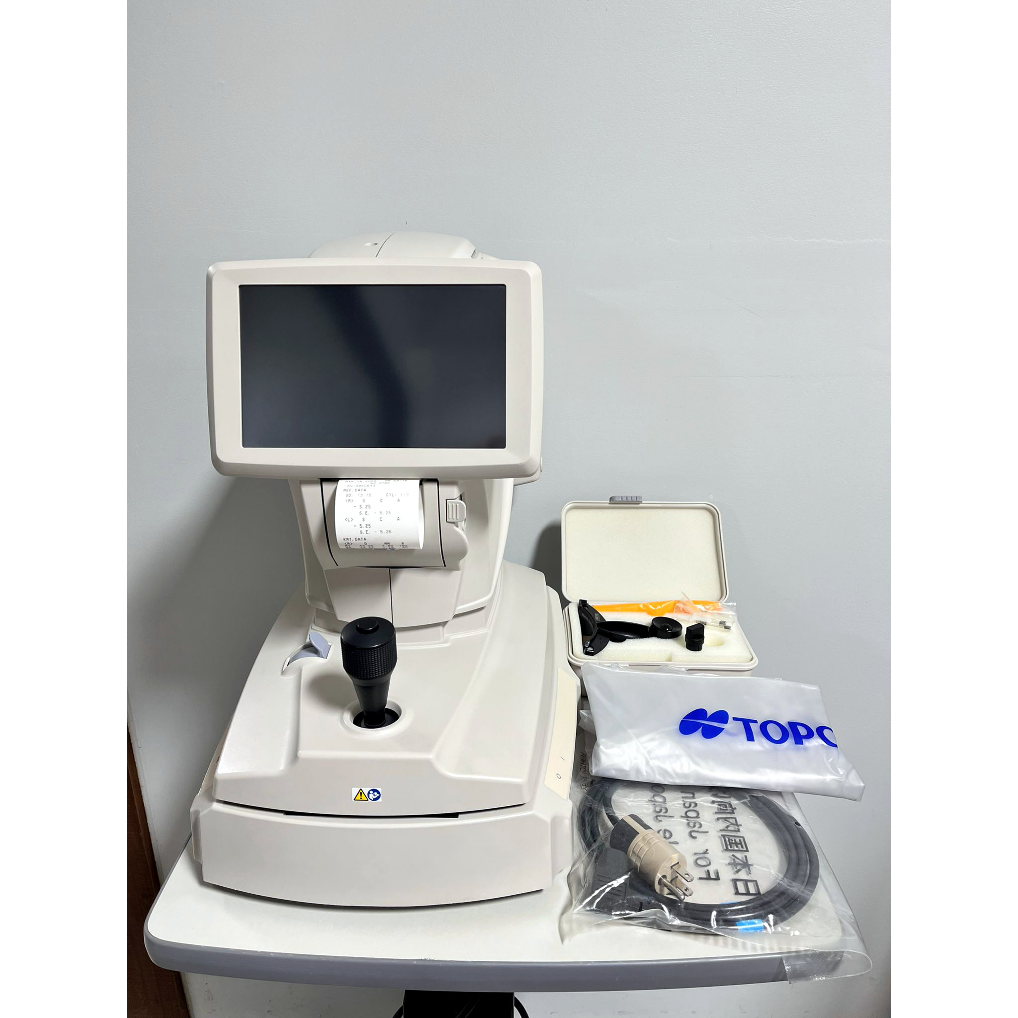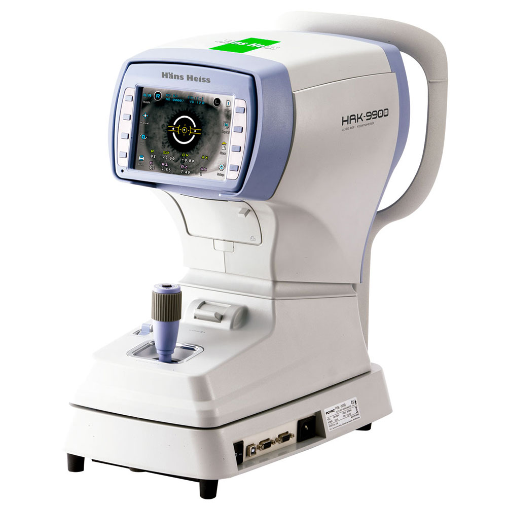
Hans Heiss HRK-9900 Autorefractor Keratometer
Refractor & Keratometry Measurement
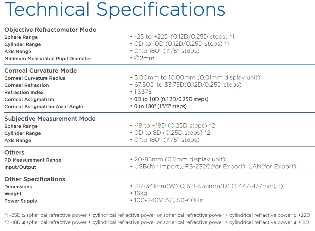
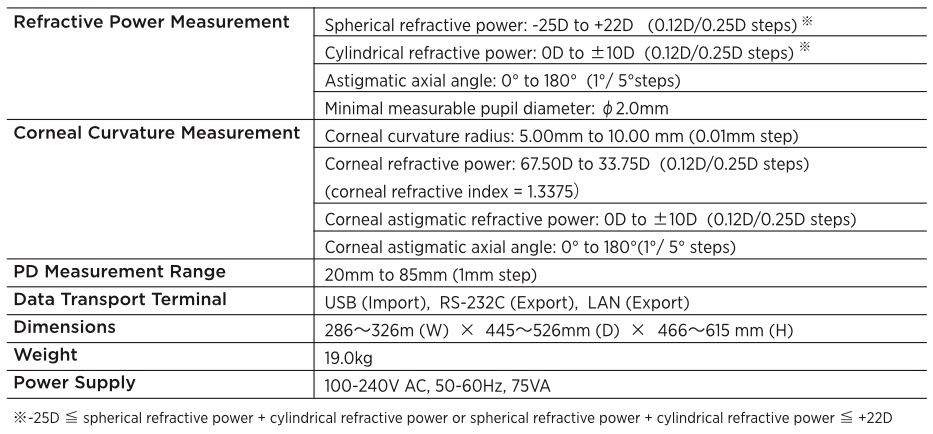
The Hans Heiss HRK-9900 provides an extensive dioptric measurement range (-25D to +22D), and the radius of curvature for keratometry is 5.0mm to 10.2mm.
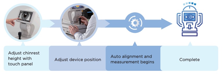
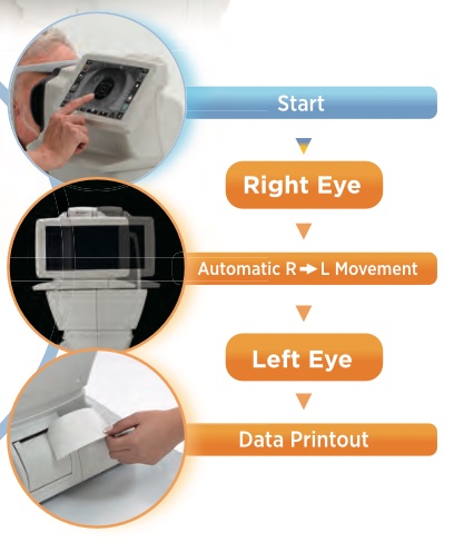
Easy alignment and friendly operation allows you to measure the refraction and keratometry in sequence, and the results can be validated immediately.
Owing to its easy alignment and friendly operation you can measure the refraction and keratometry in a sequence, the results can be checked all at once.
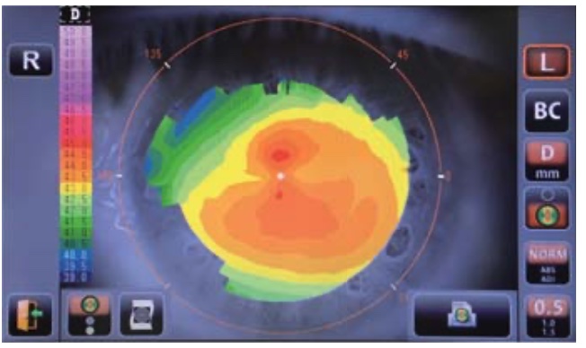
Peripheral Keratometry
Peripheral corneal curvatures can be measured by having the examinee look at the peripheral eye fixation lamps.
Measuring the corneal periphery will help you examine irregular astigmatism, and also determine a better fitting for a contact lens
Intuitive Diameter Measurement
Using the freeze function, measurement of the diameter of the cornea, pupil or hard contact lenses worn by the patient can be performed.
By simply touching and dragging the screen with your finger measurements are exactly calculated.
IOL Measurement
When refraction results in an error reading due to an intraocular lens or cataract the measurement can be performed with the IOL icon switched ‘ON’ in the Potec PRK 7000
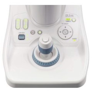
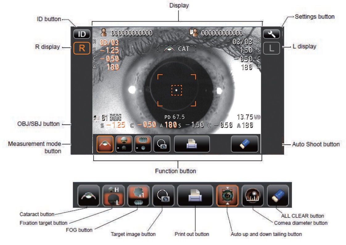
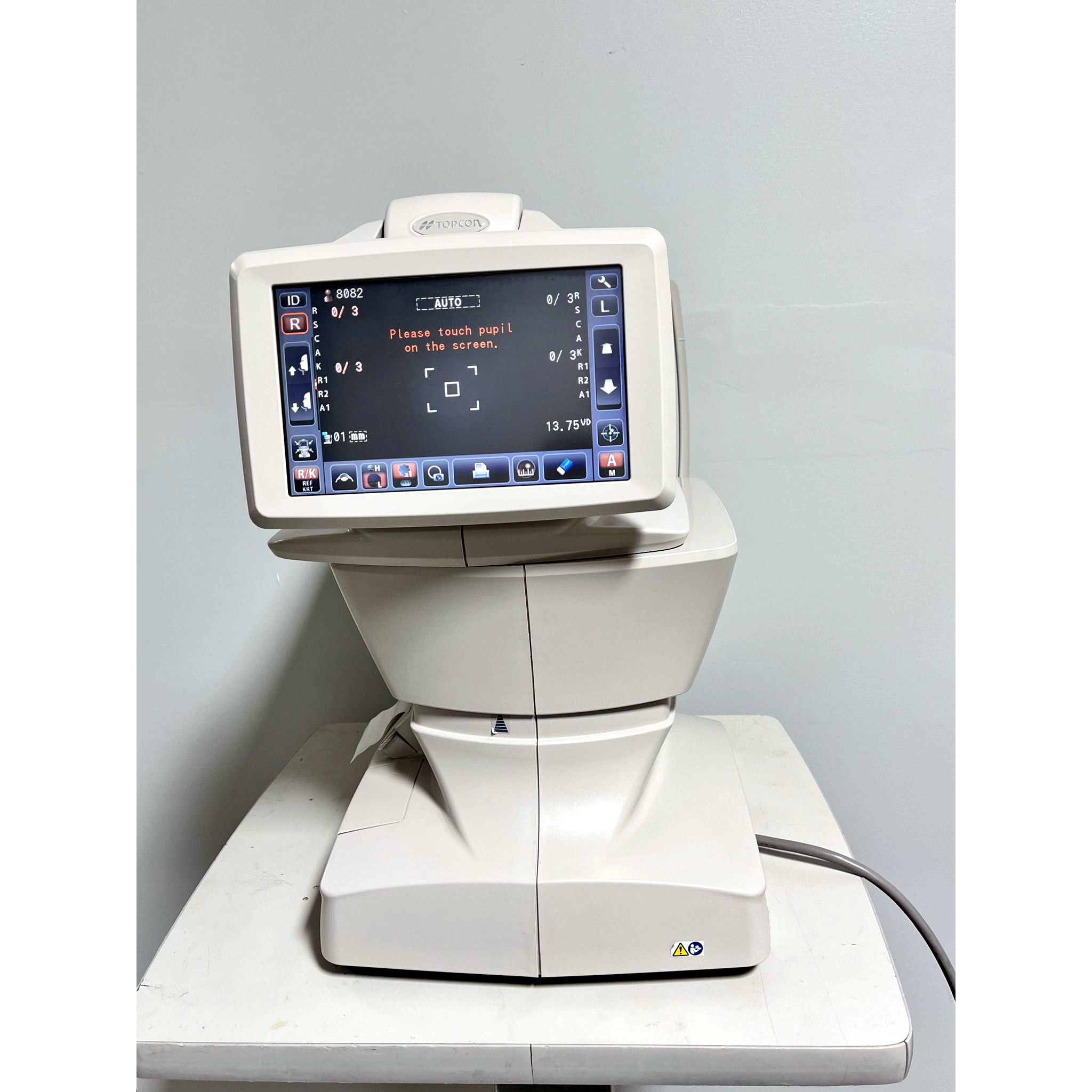
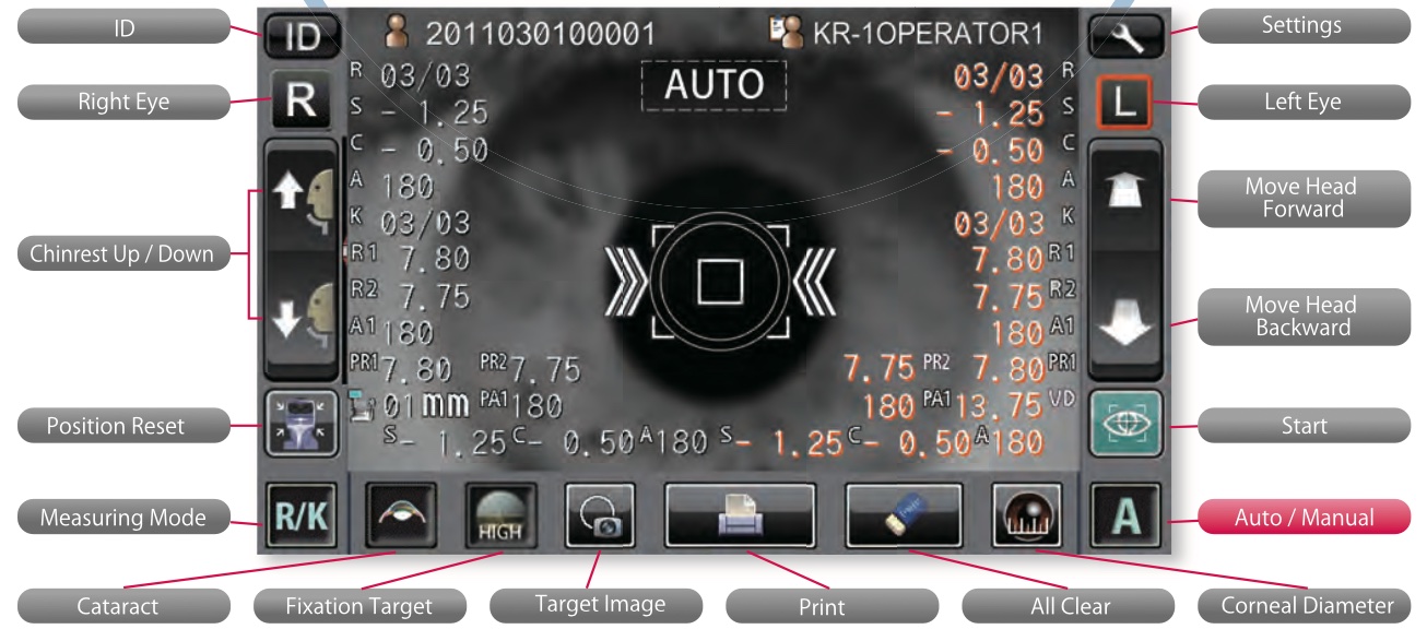
TFT-LCD with Touch Screen and Convenient control
By adopting high resolution VGA TFT-LCD with Touch Screen function, we virtually removed all input keys except the measurement button on the Joystick and Chinrest control switches.
Tests are performed rapidly and conveniently by simply pressing a button or the icon indicated on the screen. Also, user can use the Key button as convenient function.
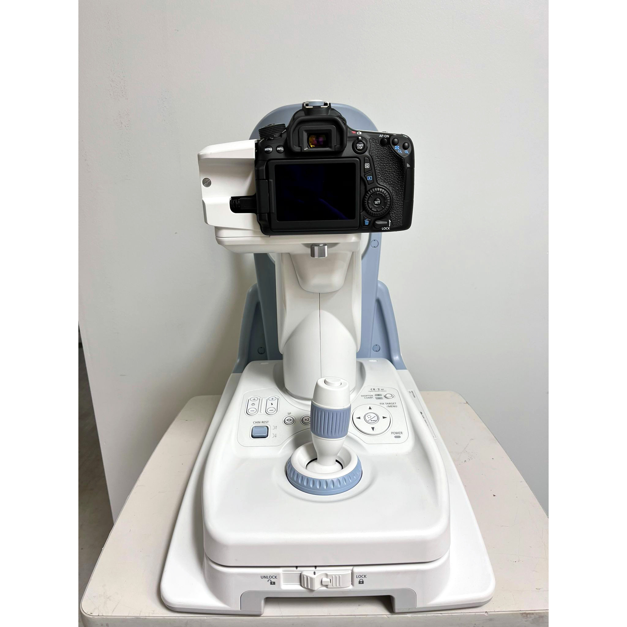
Motorized Chinrest
The chinrest is motorized with the use of a switch conveniently positioned for the operator. This makes for simple adjustment of the chinrest from patient to patient.
There is the motorized chinrest which makes user easy to control by the simple switch pressing up and down.
Interface
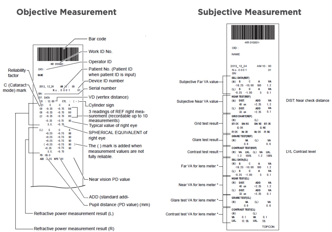
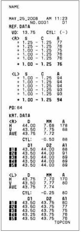
Instant Display Data
By simply touching the icon on the screen results of up to 10 measurements stored in memory can be viewed or printed by the built in thermal auto cut-off printer.
Interactive SETUP Change
Simply touch the icon on the screen and changed settings can be seen. The touch screen supports interactive setting changes quickly and conveniently saving user time innovatively.
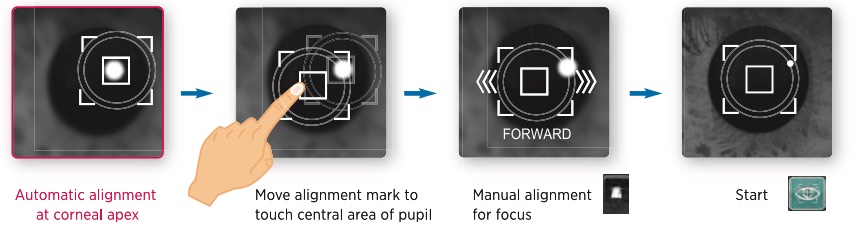
Assistant measurement function
The Hans Heiss HRK-9900 provides the assistant measurement function for the eyes which have different measuring between center of miring and center of REF focus.
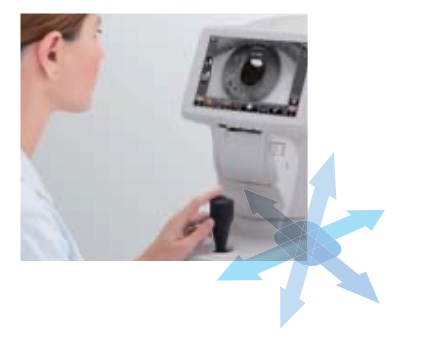
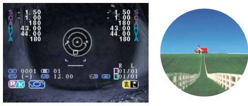
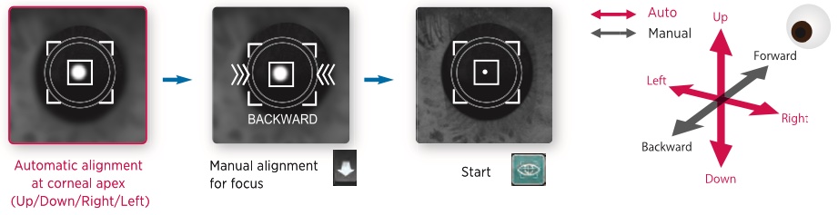
Guidance function for measuring
Hans Heiss HRK-9900 indicates the directions for focusing the center of the measuring eye and shows the conditions of focusing for the measurement convenience
Convenience
Printer with Auto Cutter Function
The Hans Heiss HRK-9900 immediately provide a complete printout of the measurement results and an auto cutter is provided for convenience. And there is the economy mode of printer which makes printer paper saved by being well arranged and amended letter size.
Data Transfer
Data can be transferred to external devices (personal computer, etc.) via an RS-232 interface. A USB interface is also provided for future compatibility. And user can send the images of patient’s eyes through USB port and have the faster S/W upgrade by the PC program which is provided as option.
Tilting LCD for user
The 90° tilting LCD provides easy control by user standing or sitting on the Potec PRK 7000
Convenience stage-lock
User can fasten the main body by simple control.
More Resources:
Potec PRK 7000 Autorefractor Keratometer