Description
What’s Included
- Carl Zeiss Cirrus 500 HD-OCT Main Unit
- Integrated Windows 10 PC (pre-installed Cirrus HD-OCT software)
- Power cable
- 90-day Digital Eye Center warranty
Condition & Warranty
Used. This Zeiss Cirrus 500 HD-OCT has been fully tested and is in excellent working order. The optical engine produces high-definition spectral-domain scans, the motorized chinrest operates smoothly, and the internal computer has been upgraded to Windows 10 for modern network security and speed. The unit shows minor signs of use as expected for pre-owned diagnostic equipment. Carefully inspected before shipping. Covered by a 90-day warranty on parts and workmanship.
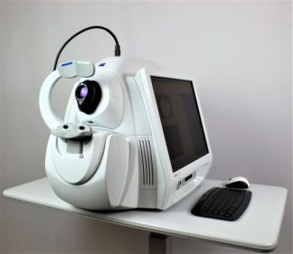
Zeiss Cirrus 500 OCT — Used HD-OCT with Windows 10
 The Zeiss Cirrus 500 OCT is the gold-standard spectral-domain optical coherence tomography system for glaucoma and retinal disease management. Powered by Zeiss’ proprietary algorithms, this Zeiss Cirrus 500 delivers high-definition cross-sectional imaging at 27,000 A-scans per second — capturing the Macular Cube 512×128 and Optic Disc Cube 200×200 protocols that clinicians rely on for repeatable, trustworthy data. This specific unit has been upgraded to a Windows 10 operating environment, providing modern cybersecurity compliance, faster data handling, and seamless integration with electronic medical records. It arrives ready for clinical use with the full Cirrus HD-OCT software suite pre-installed.
This zeiss cirrus 500 oct delivers Ganglion Cell Analysis (GCA) and Guided Progression Analysis (GPA) — tools that detect structural glaucomatous loss before visual field defects appear. Its age- and race-matched normative database gives immediate diagnostic confidence, while the compact single-table footprint integrates cleanly into any exam room without requiring additional infrastructure.
Research published in the National Library of Medicine confirms that spectral-domain OCT provides highly reproducible intraday measurements of macular layers in both glaucomatous and non-glaucomatous eyes, validating SD-OCT as the clinical standard for longitudinal structural monitoring.
Why Practices Choose the Zeiss Cirrus 500

- Windows 10 Operating System — Modern cybersecurity compliance, faster data processing, and stable network connectivity for HIPAA-compliant practice environments.
- Ganglion Cell Analysis (GCA) — Maps the macular ganglion cell–inner plexiform layer to detect early glaucomatous structural loss before visual field changes manifest.
- Guided Progression Analysis (GPA) — Compares longitudinal exam data using statistically validated algorithms to distinguish true disease progression from measurement variability.
- HD Macular Cube 512×128 — Captures a dense 3D data cube of the macula in seconds, enabling precise cross-sectional slicing and layer-by-layer analysis of retinal architecture.
- Optic Nerve Head (ONH) Analysis — Automated extraction of disc area, rim area, cup-to-disc ratio, and RNFL thickness with comparison to the Zeiss normative database.
- Extensive Normative Database — Age- and race-stratified RNFL and macular thickness baselines provide immediate, objective classification of patient measurements.
- Compact Tabletop Footprint — Integrates the optical head and processing computer into a single efficient unit that fits standard exam lanes without additional furniture.
- Proven Clinical Platform — Over a decade of peer-reviewed publications and clinical adoption makes Cirrus HD-OCT data universally understood by referring specialists.
Clinical Applications
- Glaucoma Diagnosis and Monitoring — High-precision RNFL, ONH, and Ganglion Cell layer thickness maps for early detection and longitudinal structural tracking.
- Macular Disease Assessment — Detailed layer segmentation for age-related macular degeneration (AMD), diabetic macular edema (DME), macular holes, and epiretinal membranes.
- Anterior Segment Imaging — With the optional anterior segment module, evaluate corneal thickness and anterior chamber structures (compatible accessory available separately).
- Pre- and Post-Operative Monitoring — Track anatomical changes before and after vitreoretinal or glaucoma surgery with repeatable, quantitative imaging.
- Diabetic Retinopathy Screening — Macular thickness maps and retinal layer analysis support early identification of diabetic retinal complications.
Who This Is For
- Comprehensive Eye Care Practices looking to add premium SD-OCT capability without the capital cost of a new system.
- Glaucoma and Retinal Specialists who need a secondary workstation running the trusted Zeiss analytics platform for overflow or satellite locations.
- Clinics Modernizing Infrastructure requiring Windows 10 compliance to meet hospital network security requirements.
- Mobile and Multi-Site Programs that benefit from a proven, fully functional OCT at a significantly reduced acquisition cost.
Technical Specifications
| Spec | Detail |
| Manufacturer | Carl Zeiss Meditec |
| Model | Cirrus HD-OCT 500 |
| Technology | Spectral-Domain OCT (SD-OCT) |
| Light Source | Superluminescent diode (SLD), 840 nm |
| Scan Speed | 27,000 A-scans per second |
| Axial Resolution | 5 µm (in tissue) |
| Transverse Resolution | 15 µm (in tissue) |
| Scan Depth | 2.0 mm (in tissue) |
| Scan Protocols | Macular Cube 512×128, Optic Disc Cube 200×200, HD 5-Line Raster |
| Analysis Tools | GPA, GCA, RNFL thickness maps, optic disc parameters, macular thickness maps |
| Fixation Target | Internal (Matrix LED) and External |
| Minimum Pupil Diameter | 2.0 mm |
| Operating System | Windows 10 Professional |
| Condition | Used |
| Warranty | 90-day warranty on parts and workmanship |
Compare Zeiss Cirrus HD-OCT Options
| Feature | Cirrus 400 | Cirrus 500 (This) | Cirrus 5000 |
| Scan Speed | 27,000 A/s | 27,000 A/s | 68,000 A/s |
| Axial Resolution | 5 µm | 5 µm | 5 µm |
| GPA | Yes | Yes | Yes |
| Ganglion Cell Analysis (GCA) | No | Yes | Yes |
| FastTrac™ Retinal Tracking | No | No | Yes |
| OCTA (AngioPlex) | No | No | No (separate model) |
| Operating System | Windows 10 | Windows 10 | Windows 10 |
| Condition | Used | Used | Refurbished |
| Warranty | 90-day | 90-day | 6 months |
| Price | $11,000 | $18,700 | $26,600 |
Looking to expand anterior segment imaging? The Zeiss Anterior Segment Premier Lenses are fully compatible with the Cirrus 500. For comprehensive visual field testing, consider pairing this OCT with the Zeiss Humphrey 710 Visual Field Analyzer.
Questions about the Zeiss Cirrus 500 OCT? Contact us for availability and shipping details.