Description
What’s Included
- Optovue iVue Spectral-Domain OCT Engine (26,000 A-scans/second, 5 µm axial resolution)
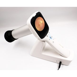
- iCam Non-Mydriatic Fundus Camera Module (5.2 MP, 45° field of view)
- Medical-grade electric elevation table (motorized vertical positioning)
- Integrated Optovue Software Suite (retinal, optic nerve, glaucoma, anterior segment protocols)
- Complete cables, accessories, and operator documentation
- 90-day warranty on parts and workmanship
Condition & Warranty
Used. Fully tested and confirmed in excellent working condition. The SD-OCT engine delivers sharp, high-resolution cross-sectional scans with consistent 5 µm axial precision. The iCam module produces crisp, high-resolution color fundus images. The electric table operates smoothly across its full vertical range. Minor cosmetic wear. Covered by a 90-day warranty on parts and workmanship.
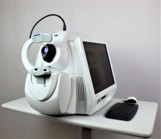
Optovue iFusion iCam — Used Combined OCT & Fundus Camera
 The Optovue iFusion iCam combines the iVue spectral-domain OCT with the iCam high-resolution fundus camera in a single diagnostic platform. At 26,000 A-scans per second and 5 µm axial resolution, the iVue SD-OCT delivers the imaging depth and speed needed for RNFL mapping, macular layer segmentation, GCC analysis, and anterior segment evaluation. The integrated iCam non-mydriatic fundus camera adds 5.2 MP, 45° color retinal photography through pupils as small as 4 mm — without requiring dilation or patient repositioning.
This Optovue iFusion iCam system eliminates the need for separate OCT and fundus imaging instruments, dramatically improving workflow efficiency and reducing examination time in high-volume screening environments. The included medical-grade electric elevation table provides motorized vertical positioning to accommodate all patient demographics without manual adjustment.
Research published in the National Library of Medicine confirms that combining optical coherence tomography with color fundus photography significantly improves glaucoma diagnostic accuracy compared to either modality alone — precisely the dual-imaging approach the Optovue iFusion iCam delivers in a single unit.
Why Practices Choose the Optovue iFusion iCam

- True Dual-Modality Integration — SD-OCT and fundus photography in a single unit without patient repositioning
- Ultra-Fast OCT Scanning — 26,000 A-scans per second with high-density retinal imaging protocols
- Exceptional Depth Resolution — 5 µm axial resolution enables precise early disease detection and layer segmentation
- Non-Mydriatic Fundus Imaging — 5.2 MP, 45° field-of-view color imaging through small pupils without dilation
- Comprehensive Analysis Protocols — Retinal thickness, RNFL, optic nerve head, GCC segmentation, anterior segment
- Integrated Electric Table — Motorized vertical positioning for all patient demographics, included with the system
- Normative Comparison Databases — Built-in glaucoma risk stratification and longitudinal trend tracking
Clinical Applications
- Glaucoma Screening & Management — RNFL thickness mapping with simultaneous optic nerve fundus documentation
- Age-Related Macular Degeneration — High-resolution retinal layer assessment for early detection and progression monitoring
- Diabetic Macular Edema — Precise retinal thickness quantification co-registered with color fundus documentation
- Diabetic Retinopathy — Automated layer segmentation, thickness trending, and photo documentation in one visit
- Anterior Segment Evaluation — Corneal pachymetry, anterior chamber depth measurement, lens opacity assessment
- Optic Nerve Disorders — GCC segmentation to differentiate glaucomatous from non-glaucomatous neuropathy
Who This Is For
- Comprehensive Ophthalmology & Optometry Practices — Advanced dual-modality imaging without expanding clinic footprint
- High-Volume Screening Suites — Rapid unified documentation for diabetic retinopathy and glaucoma programs
- Specialty Retinal & Glaucoma Clinics — Objective structural imaging with co-registered fundus photos for diagnosis and follow-up
- Research & Academic Institutions — Multi-protocol retinal disease studies requiring both structural and photographic data
Technical Specifications
| Specification | Detail |
| System Name | Optovue iFusion (iVue SD-OCT + iCam Fundus Camera) |
| OCT Technology | Spectral-Domain (SD-OCT) |
| Scan Speed | 26,000 A-scans per second |
| Axial Resolution | 5 µm (in tissue) |
| Transverse Resolution | 15 µm at retina |
| Scan Beam Wavelength | 840 nm ± 10 nm |
| Fundus Camera | iCam — 5.2 MP, 45° field of view, non-mydriatic |
| Minimum Pupil Size | 4.0 mm (non-mydriatic operation) |
| Working Distance (Retina) | 21.2 mm |
| Working Distance (Cornea) | 16.6 mm |
| Motorized Focus Range | -15D to +10D |
| Scan Protocols | Retinal, Glaucoma (RNFL/GCC), Anterior Segment, Macula |
| Electric Table | Integrated, motorized vertical positioning |
| Condition | Used (fully tested, excellent working condition) |
| Warranty | 90-day parts and workmanship |
Compare Diagnostic Imaging Options
| Feature | Optovue iFusion iCam | Zeiss Cirrus 500 | Zeiss Clarus 500 |
| Type | SD-OCT + Fundus Camera (combined) | SD-OCT only | Ultra-widefield camera only |
| OCT Technology | SD-OCT, 26,000 A-scans/sec, 5 µm | SD-OCT, 27,000 A-scans/sec, 5 µm | None |
| Fundus Imaging | 5.2 MP, 45° non-mydriatic | None | 133°–200° multi-modal (True Color, FAF, IR) |
| Electric Table | Included | Not included | Not included |
| Condition | Used | Refurbished | Used |
| Warranty | 90 days | 6 months | 90 days |
| Price | $18,700 | $20,900 | $27,500 |
Also consider: Zeiss Cirrus 500 OCT for standalone SD-OCT, or the Zeiss Clarus 500 for ultra-widefield retinal imaging.
Support & Resources
Every Optovue iFusion iCam system includes complete documentation and technical support. We provide post-sale guidance on setup, operation, and optimization. For advanced training, we can connect you with Optovue’s authorized service network. Contact us with any questions.