Description
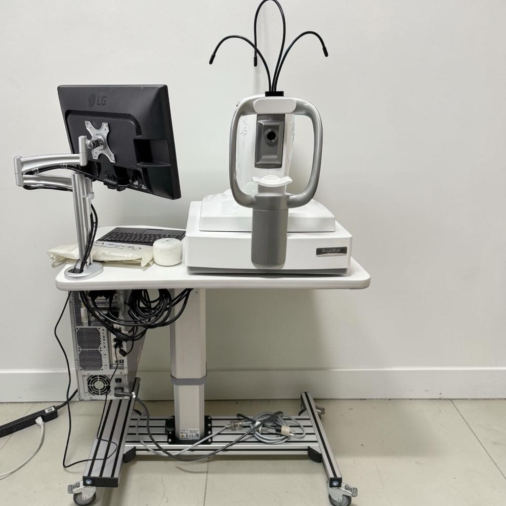
What’s Included
-
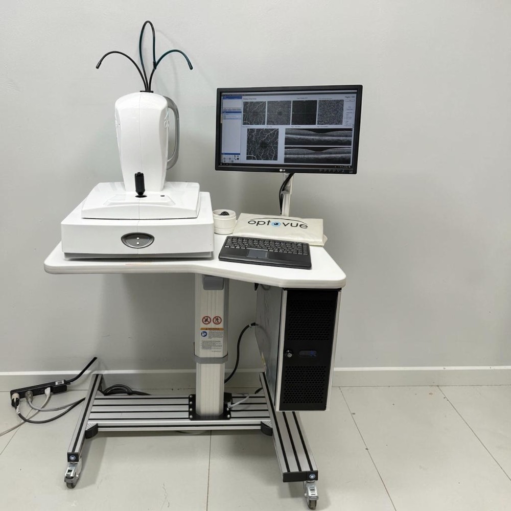
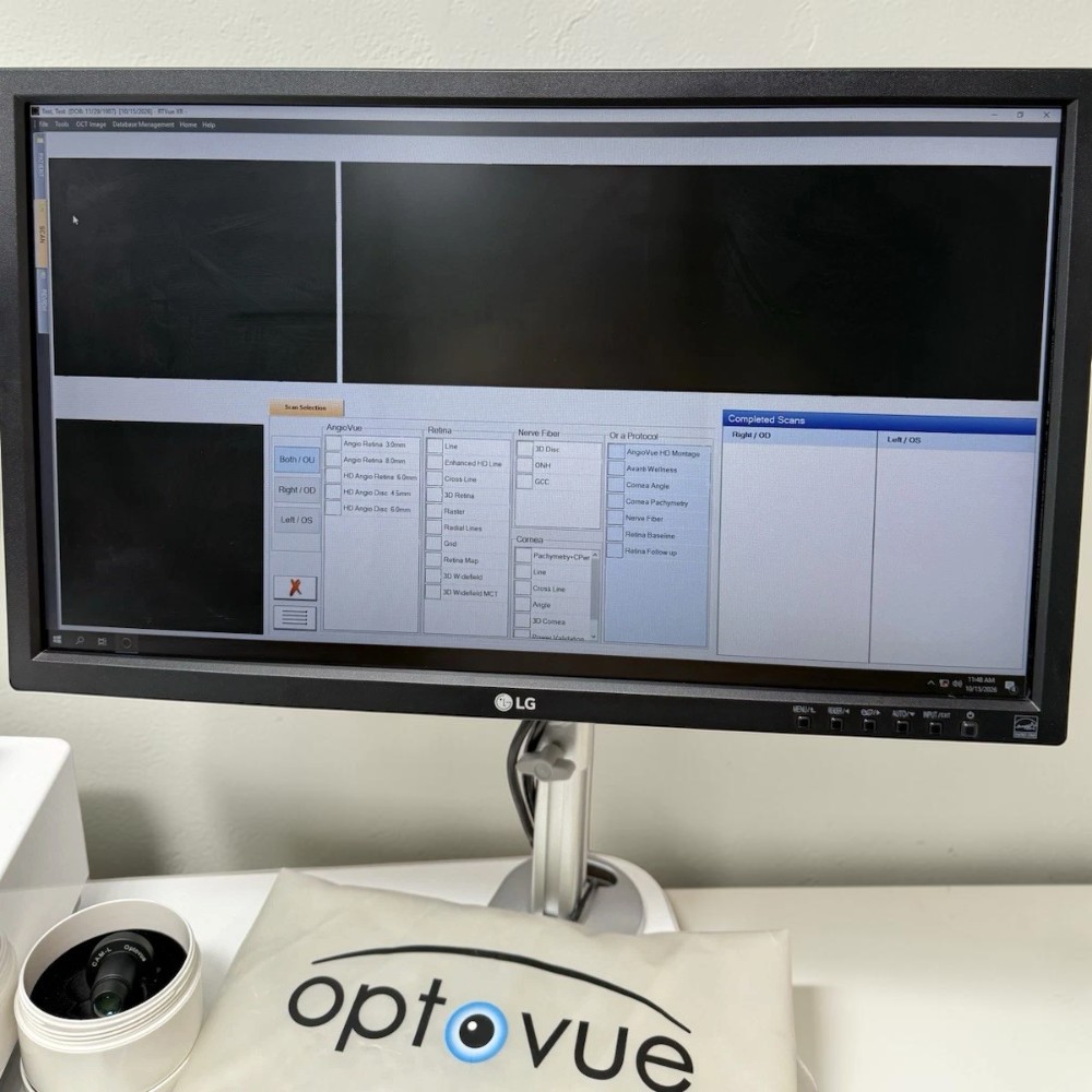
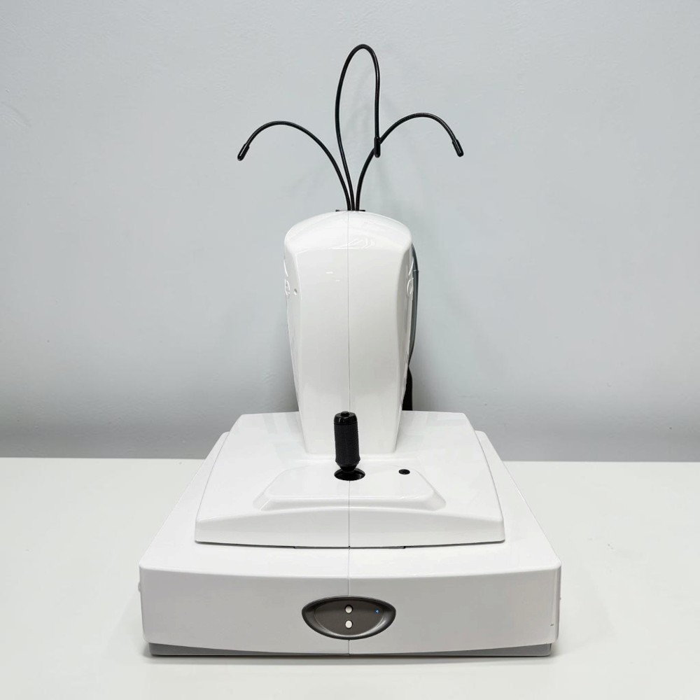
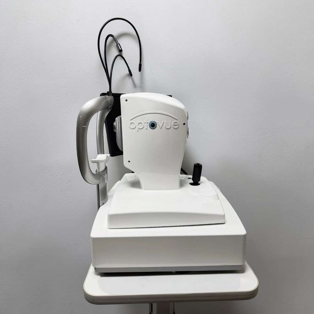
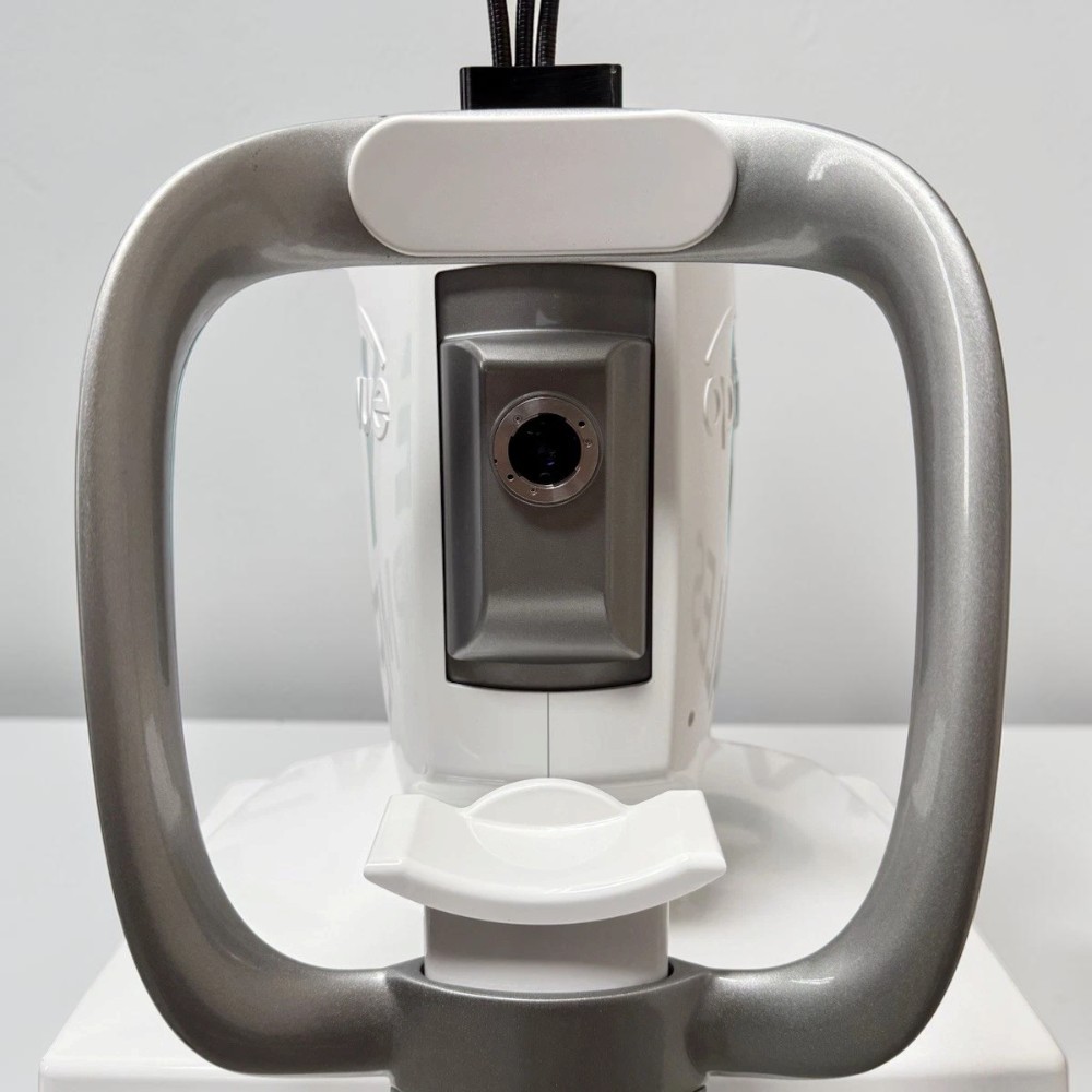
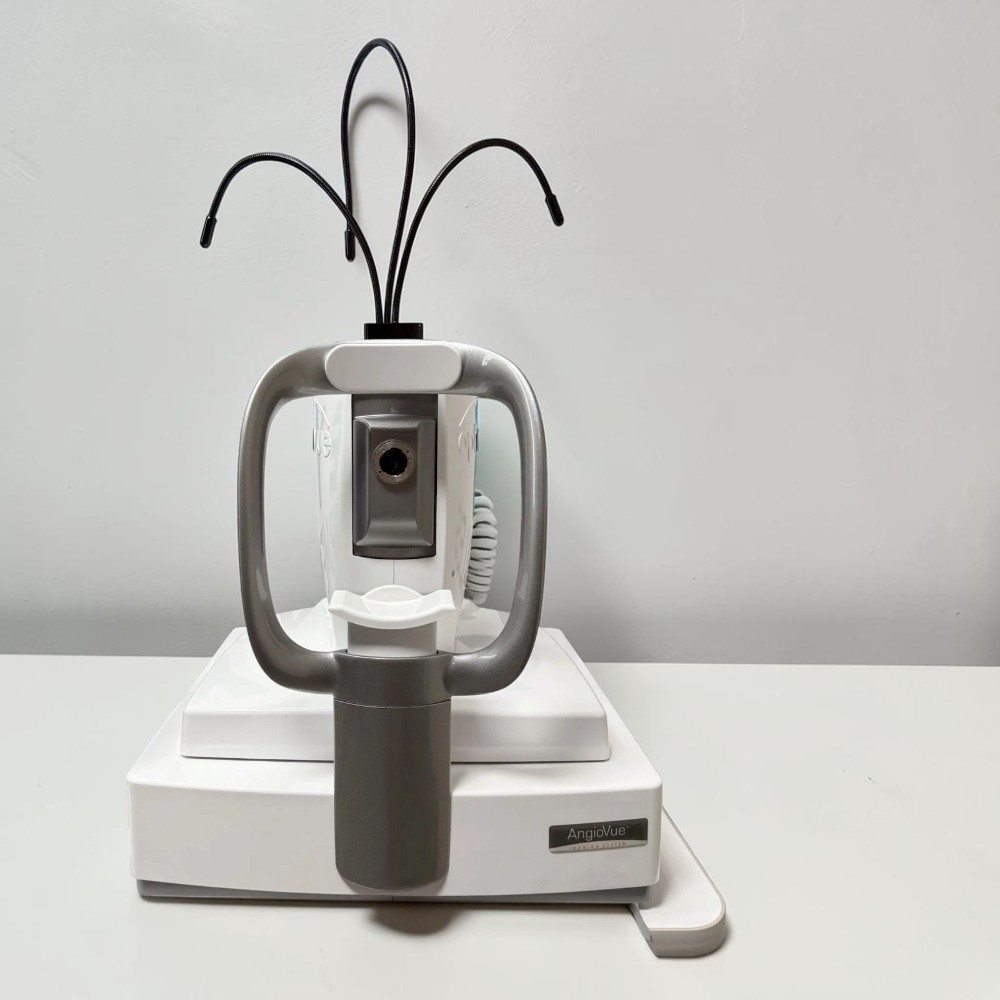
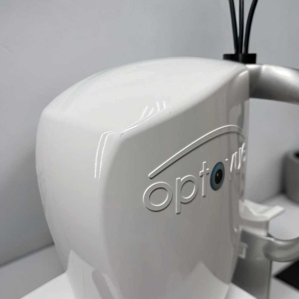
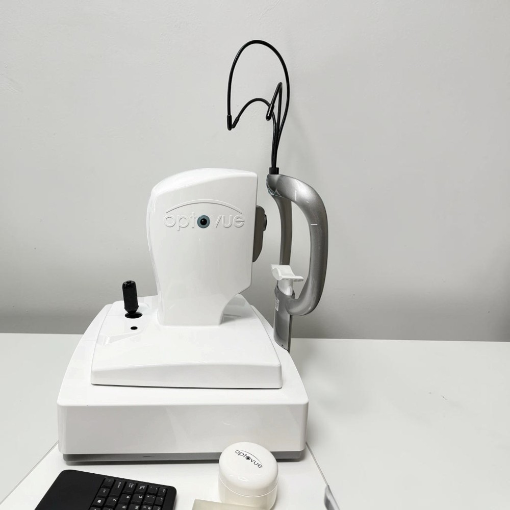
Optovue AngioVue OCTA Imaging System
-
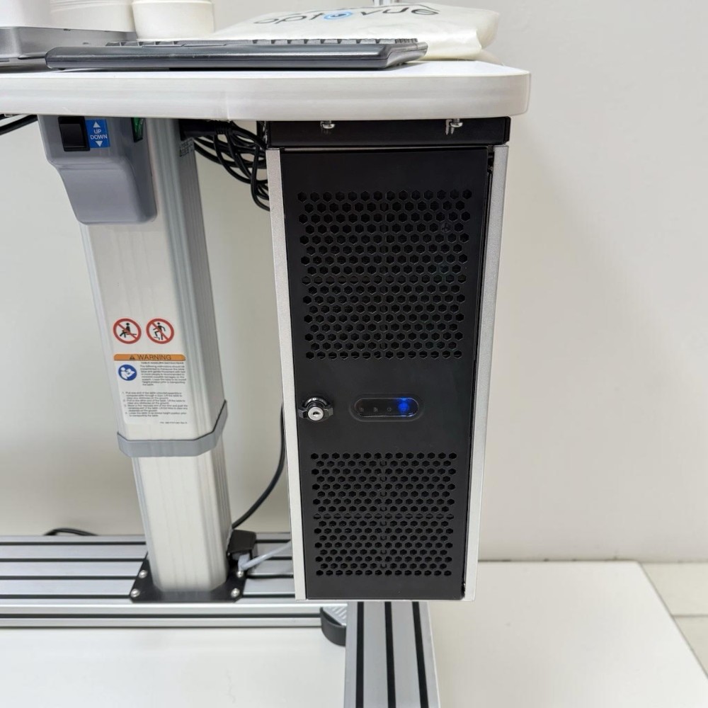
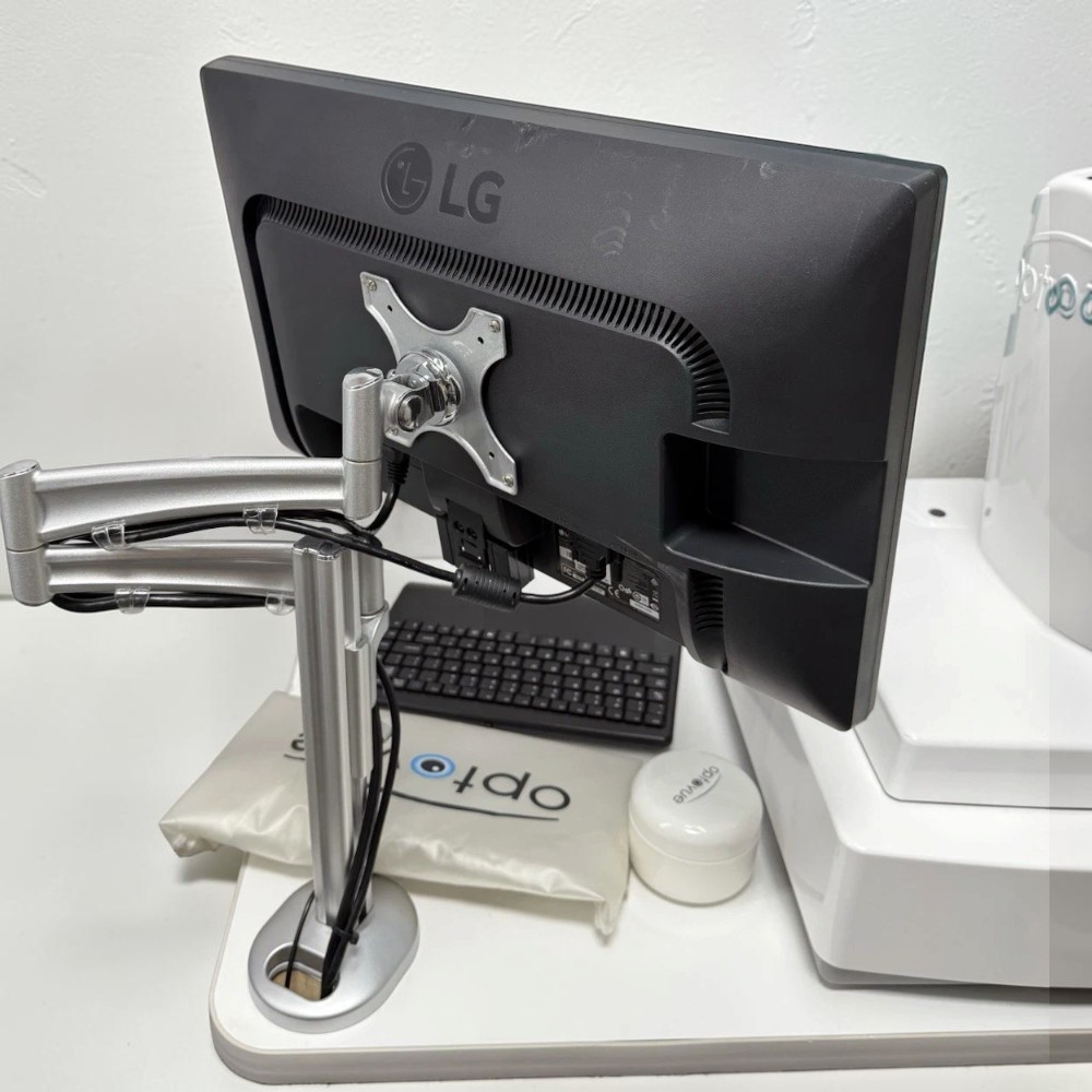
Electric table
-
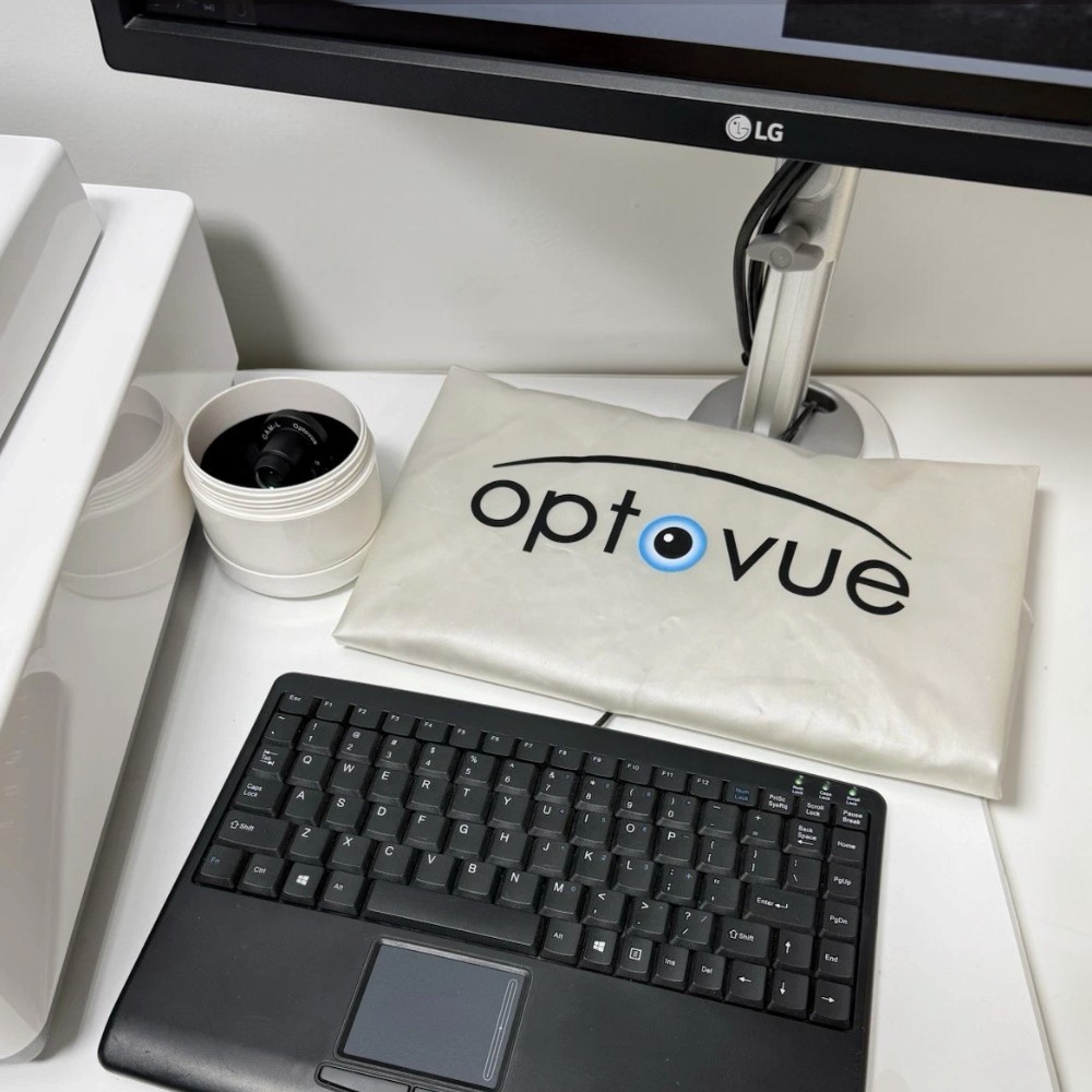
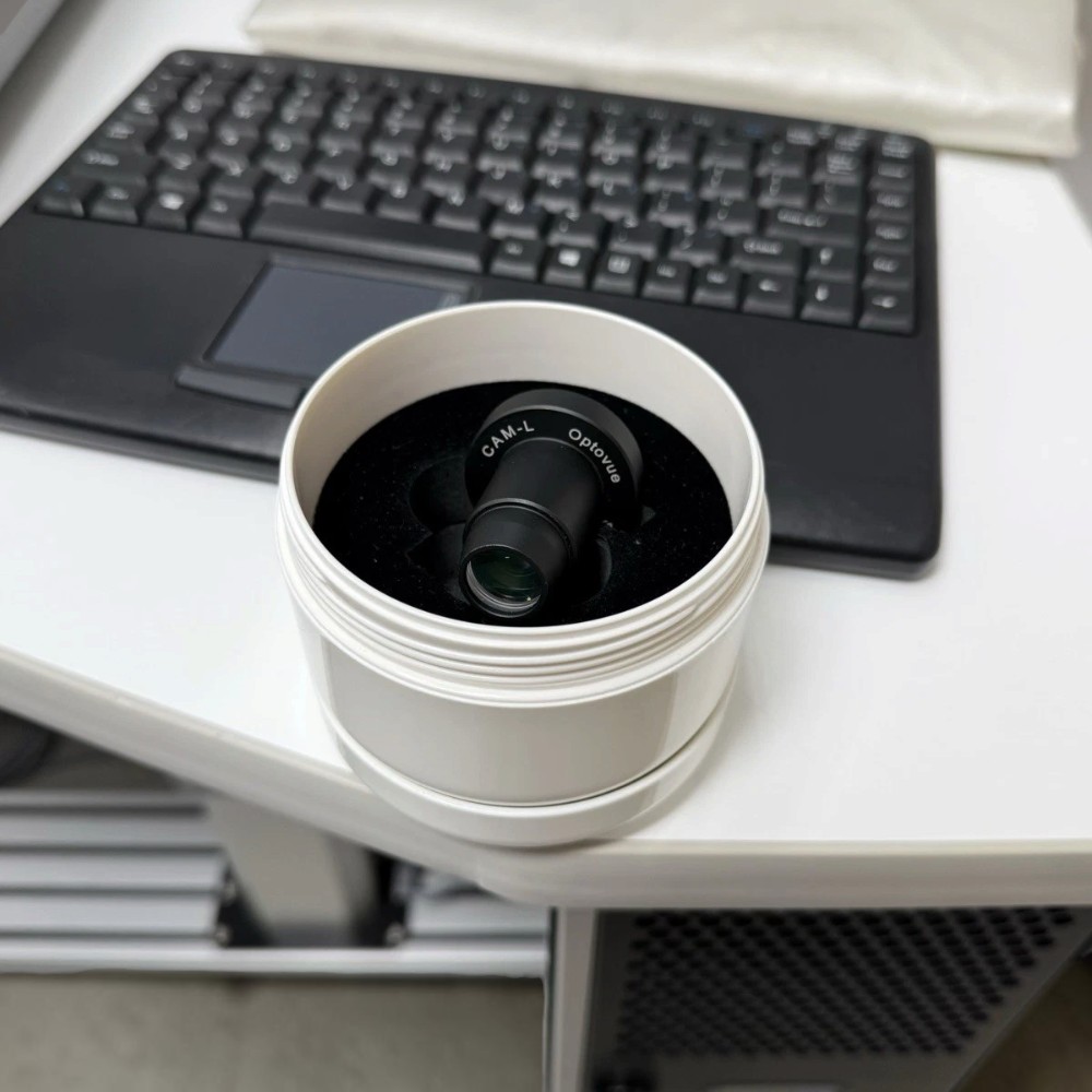
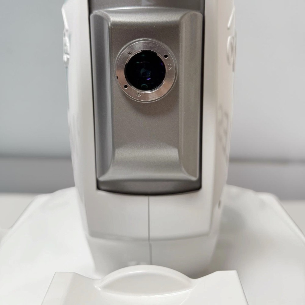
CAM-L lens
-
Power cable
-
Dust cover
-
90-day Digital Eye Center warranty
Condition & Warranty
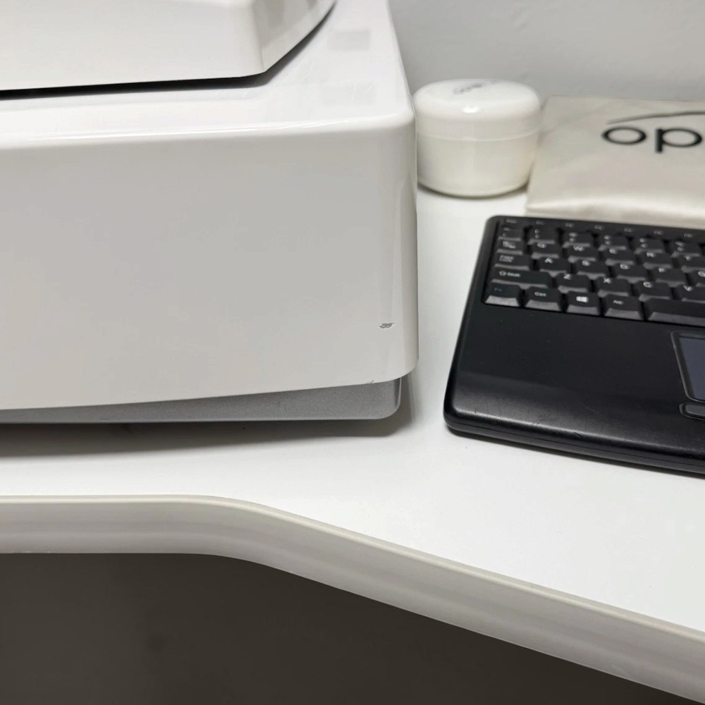
Used. The Optovue AngioVue Imaging System has been tested and in excellent working condition. The unit has been cleaned and is ready for use upon receipt, according to the listing. Cosmetic signs of previous use may be present. Covered by a 90-day Digital Eye Center warranty on parts and workmanship.
Optovue AngioVue OCTA Imaging System — Optical Coherence Tomography Angiography
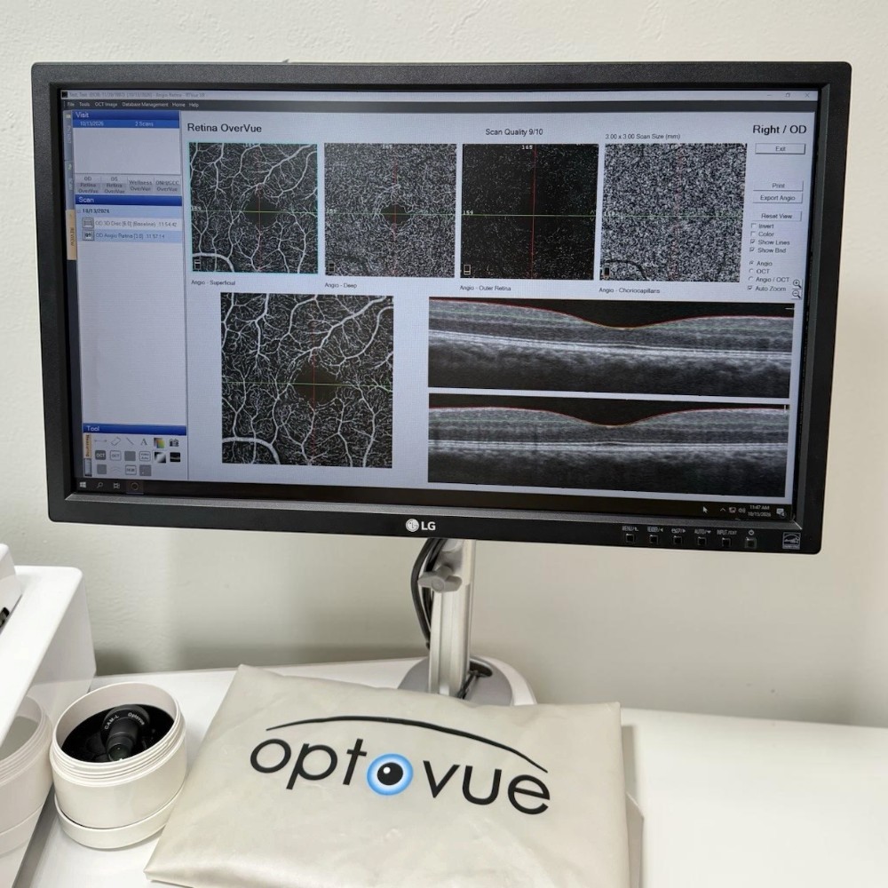
The Optovue AngioVue is an ophthalmic imaging system that combines spectral-domain optical coherence tomography (SD-OCT) with optical coherence tomography angiography (OCTA). It produces cross-sectional images of retinal structures and visualizations of retinal and choroidal blood vessels without requiring intravenous dye for angiographic imaging.
AngioVue uses Optovue’s Split-Spectrum Amplitude-Decorrelation Angiography (SSADA) technology to detect blood-flow signals and generate detailed vascular maps. The system is designed to support assessment of retinal microvasculature, macular conditions, and optic nerve head circulation.
The AngioVue platform is associated with the RTVue XR Avanti SD-OCT system. Manufacturer documentation lists an OCT scanning speed of 70,000 A-scans per second, approximately 5-micron axial optical resolution, and multiple OCTA scan sizes. The precise software version, licensing, and configuration of this used unit should be established from the unit itself and its included software.
Why Practices Choose the Optovue AngioVue
-
Noninvasive Vascular Imaging — Visualizes retinal microvasculature without intravenous contrast dye.
-
High-Speed OCT Acquisition — Captures up to 70,000 A-scans per second.
-
Detailed Retinal Imaging — Provides approximately 5-micron axial optical resolution and 15-micron transverse optical resolution.
-
Multiple OCTA Scan Sizes — Supports retinal imaging protocols of 3 × 3 mm, 6 × 6 mm, and 8 × 8 mm, depending on configuration.
-
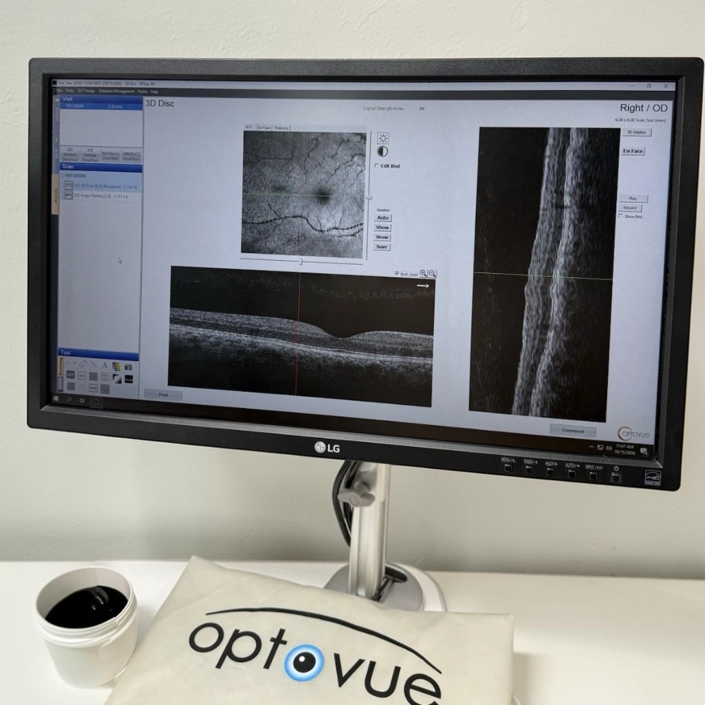
Optic Disc Analysis — Supports OCTA imaging of the optic nerve head and peripapillary vascular structures.
-
Structural OCT and Angiography — Combines cross-sectional retinal imaging with vascular visualization.
-
Integrated Workstation — The listed package includes an electric table, CAM-L lens, power cable, and dust cover.
Clinical Applications
-
Retinal Vascular Assessment — Visualization of retinal capillary networks and vascular abnormalities.
-
Diabetic Retinopathy Evaluation — Assessment of retinal microvascular changes associated with diabetes.
-
Age-Related Macular Degeneration (AMD) — Evaluation of retinal and choroidal vascular changes.
-
Macular Disease Assessment — Imaging of vascular structures in the macular region.
-
Retinal Vein Occlusion — Evaluation of retinal perfusion and vascular changes.
-
Glaucoma Assessment — Evaluation of optic nerve head and peripapillary vascular structures.
-
Choroidal Neovascularization Assessment — Visualization of abnormal vascular networks when detectable with the appropriate OCTA protocol.
-
Longitudinal Monitoring — Comparison of structural OCT and OCTA images across clinical visits.
Who This Is For
-
Retina Specialists evaluating retinal vascular disorders and macular diseases.
-
Ophthalmologists requiring structural OCT and OCT angiography.
-
Glaucoma Specialists assessing optic nerve head structure and vascular features.
-
Multispecialty Eye Clinics seeking advanced retinal imaging capabilities.
-
Ophthalmic Research Centers studying retinal and choroidal microvasculature.
Technical Specifications
| Specification | Detail |
|---|---|
| Manufacturer | Optovue |
| System | AngioVue OCTA |
| OCT Platform | RTVue XR Avanti SD-OCT |
| Imaging Modalities | Spectral-domain OCT and OCT angiography (OCTA) |
| OCT Light Source | Approximately 840 nm |
| OCT Scanning Speed | 70,000 A-scans per second |
| Optical Axial Resolution | Approximately 5 µm |
| Digital Pixel Sampling | 3 µm |
| Optical Transverse Resolution | Approximately 15 µm |
| OCT Axial Imaging Depth | 2–3 mm, depending on scan protocol |
| AngioVue Imaging Volume | 304 × 304 A-scans for standard scans; 400 × 400 A-scans for HD scans |
| OCTA Acquisition Time | Approximately 3 seconds per imaging volume |
| Retinal Scan Sizes | 3 × 3 mm, 6 × 6 mm HD, 8 × 8 mm, depending on configuration |
| Optic Disc Scan Sizes | 4.5 × 4.5 mm HD and 6 × 6 mm HD, depending on configuration |
| Field of View | 12 × 9 mm |
| Minimum Pupil Diameter | Approximately 2.3 mm |
| Angiography Algorithm | Split-Spectrum Amplitude-Decorrelation Angiography (SSADA) |
| Computer Operating System | Windows 64-bit; exact supported version depends on installed software |
| Minimum RAM | 8 GB |
| Recommended RAM | 16 GB |
| Recommended Processor | Intel i7, 3 GHz or higher |
| Minimum Hard Drive Availability | 50 GB |
| Network Bandwidth | 1 Gbps or higher |
| Table Width | 950 mm (37.4 in) |
| Table Depth | 600 mm (23.6 in) |
| Adjustable Table Height | 695–995 mm (27.4–39.2 in) |
| Included Accessories | Electric table, CAM-L lens, power cable, dust cover |
| Condition | Used |
| Warranty | 90-day Digital Eye Center warranty on parts and workmanship |