Description
This exact unit sold recently — but we specialize in sourcing refurbished ophthalmic equipment. Typical turnaround is 2–4 weeks once we confirm availability with our partners. Request a free quote with no deposit required.
Request Availability → Free quote · No obligation · 90-day warranty when shipped
What’s Included
- Topcon Maestro 1 OCT all-in-one combo (OCT + non-mydriatic color fundus)
- Workstation / PC with Maestro software
- Power cabling (as photographed)
- 6-month Digital Eye Center warranty
Condition & Warranty
Refurbished. Tested and refurbished before listing — 50,000 A-scans/sec spectral-domain OCT engine verified, 12×9 mm 3D macula and disc cube acquisition confirmed, non-mydriatic color fundus capture validated, auto-alignment + auto-focus + auto-capture confirmed, FastMap report generation verified, calibration check completed. Covered by a 6-month warranty on parts and workmanship.
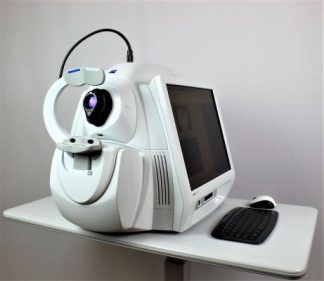
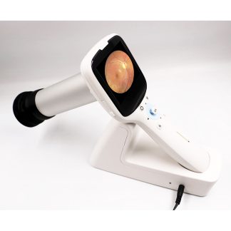
Topcon Maestro 1 — Refurbished All-in-One OCT + Non-Mydriatic Color Fundus Combo
 The Topcon Maestro 1 OCT is Topcon’s all-in-one combo platform — spectral-domain OCT plus non-mydriatic color fundus camera in a single instrument with single-button auto-acquisition of both modalities. This refurbished topcon maestro ships ready for the standard combo workflow: one push, both OCT and fundus image captured in seconds with auto-alignment, auto-focus, and auto-capture.
The Maestro’s headline advantage is workflow efficiency — instead of moving a patient between separate OCT and fundus-camera stations, the Maestro completes both acquisitions in one chair-side session. The 12×9 mm 3D cube simultaneously captures macula and optic disc data, supporting both retina and glaucoma workups from a single scan. Ideal for high-volume optometry and screening clinics.
According to a National Library of Medicine-indexed study (Indian J Ophthalmol, 2026), ophthalmic-instrument repeatability depends heavily on operator-instrument interaction — supporting the standardized auto-acquisition workflow the Maestro was designed to deliver.
Why Practices Choose the Maestro
 All-in-one combo — OCT + non-mydriatic color fundus in one instrument.
- Auto-alignment + auto-focus + auto-capture — single-button workflow.
- 50,000 A-scans/sec — Topcon spectral-domain OCT speed.
- 12×9 mm 3D cube — macula + disc in one scan.
- Non-mydriatic color fundus — no dilation required.
- FastMap reporting — Topcon glaucoma + retina analysis.
- Compact footprint — fits standard exam-lane configurations.
- Refurbished tier with 6-month DEC warranty — substantial savings vs new.
Clinical Applications
- Glaucoma + retina combo workup — single-scan optic disc + macula coverage.
- Diabetic retinopathy screening — fundus + OCT macular thickness in one session.
- AMD diagnosis and follow-up — non-myd color fundus + OCT macular cube.
- Glaucoma RNFL surveillance — optic disc cube within the 12×9 mm field.
- High-volume optometry pretest — fast auto-capture workflow.
- Tele-retina screening — combined OCT + fundus referral packets.
Who This Is For
- Optometry practices wanting OCT + fundus camera in one footprint.
- Screening clinics needing high-throughput auto-capture workflow.
- Multi-doctor practices standardizing on Topcon imaging.
- Practices with limited exam-lane space needing combo functionality.
- Buyers comparing Maestro vs separate Topcon NW-series fundus + Cirrus/Avanti OCT.
Technical Specifications
| Specification | Detail |
| Type | All-in-one SD-OCT + non-mydriatic color fundus camera |
| Scan speed | 50,000 A-scans/sec |
| Scan field | 12×9 mm 3D cube (macula + disc) |
| Fundus modality | Color, non-mydriatic |
| Automation | Auto-alignment, auto-focus, auto-capture |
| Reporting | FastMap glaucoma + retina analysis |
| Manufacturer | Topcon (Japan) |
| Condition | Refurbished — tested before listing |
| Warranty | 6-month DEC warranty on parts and workmanship |
Compare OCT Options
Where this Topcon Maestro fits in DEC’s OCT inventory:
| Model | Combo | OCT-A | Brand | Price | Notes |
| Topcon Maestro 1 | Yes (OCT + fundus) | No | Topcon | $17,000 | All-in-one combo — this listing |
| Topcon 3D OCT-2000 | Yes (OCT + fundus + FA) | No | Topcon | $15,600 | Combo + FA capability |
| Zeiss Cirrus 6000 | OCT only | AngioPlex 12×12 mm | Zeiss | $52,000 | Flagship Zeiss OCT-A — Refurbished |
| Optovue Avanti XR | OCT only | AngioVue 12×9 mm | Optovue | $38,500 | Widefield OCT-A — Refurbished |
| Zeiss Cirrus 5000 OCT | OCT only | Optional | Zeiss | $26,600 | In stock — Used |
| Zeiss Cirrus 4000 | OCT only | No | Zeiss | $15,400 | In stock — Used |
| Optovue RTVue-100 | OCT + anterior | No | Optovue | $12,500 | In stock — Refurbished anterior |
Similar OCTs In Stock Now
Need a comparable oct available today? These units are in stock and ready to ship with a 90-day Digital Eye Center warranty.


Prefer to wait for the Topcon Maestro 1 OCT – Refurbished? Request availability and we’ll source one for you.
Questions about the Topcon Maestro? Contact us for current availability and shipping details.