Description
What’s Included
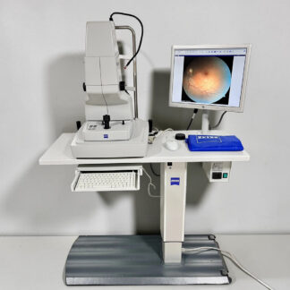
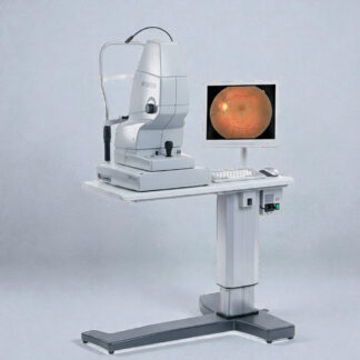
- Carl Zeiss VISUCAM PRO NM fundus camera (color non-mydriatic, basic NM model)
- Lenovo capture computer (per existing listing)
- Electric Table (per listing title)
- Power cable
- 90-day Digital Eye Center warranty
Condition & Warranty
Used. Tested before listing — color fundus capture verified at both 45° and 30° fields, ø3.3 mm small-pupil mode confirmed, RGB channel-separated capture verified, electric table movement confirmed. Cleaned. Covered by a 90-day warranty on parts and workmanship. Note: this is the basic NM model — Fluorescein Angiography (FA), Fundus Autofluorescence (FAF), and Indocyanine Green (ICG) modules are NOT included on this configuration (those are on the NMFA / NMFA-FAF-ICG variants).
Visucam PRO NM — Used Color Non-Mydriatic Fundus Camera with Electric Table
 The Visucam PRO NM is the entry-tier fundus camera in the Zeiss VISUCAM family — non-mydriatic color fundus photography on the same ZEISS Telecentric Optics platform as the higher-tier NMFA and NMFA-FAF-ICG variants, but without the angiography modules. This used visucam pro nm ships on its electric table for immediate clinical deployment.
The system uses a 5.0 MP digital sensor with ZEISS Telecentric Optics, paired with a 45° / 30° dual field of view and a minimum pupil diameter of ø3.3 mm for non-mydriatic capture. Acquisition modes include color fundus and channel-separated green / red / blue images (as separate photos or as RGB layers of a color photo) — useful for vessel and RNFL review without the cost of full FA capability.
According to a National Library of Medicine-indexed multicenter study (Int J Gen Med, 2023), non-mydriatic fundus cameras deliver high diagnostic sensitivity for referral-warranted diabetic retinopathy in primary-care settings — exactly the workflow the Visucam PRO NM was built around.
Why Practices Choose the Visucam PRO NM
 5.0 MP Zeiss Telecentric Optics — premium Zeiss optical platform shared with the higher NMFA tiers; produces clean, high-contrast color fundus images.
- Dual 45° / 30° field — full-angle 45° captures the posterior pole; 30° digital crop highlights disc and macula detail.
- Non-mydriatic ø3.3 mm pupil — undilated capture for primary-care DR screening and routine optometry workflows.
- RGB channel-separated capture — green, red, and blue images as separate photos or as RGB layers; useful for vessel inspection and RNFL review.
- DICOM conformant / FORUM compatible — fits cleanly into a Zeiss imaging stack alongside Cirrus OCT and Humphrey perimeters.
- Electric table included — height-adjustable powered base ships with the unit, simplifying patient positioning across exam lanes.
- Color-only entry tier — significantly lower cost than NMFA / FAF / ICG variants for clinics that don’t perform in-house angiography.
- Same ergonomics as NMFA siblings — staff trained on a NM transitions seamlessly to NMFA upgrade if/when angiography is added.
Clinical Applications
- Diabetic retinopathy screening — non-mydriatic 45° color capture for chronic-disease primary-care, optometry, and tele-retinal programs.
- Macular degeneration follow-up — true-color images document drusen, geographic atrophy, and pigmentary changes over time.
- Glaucoma disc documentation — color disc photos for cup/disc ratio tracking.
- Hypertensive retinopathy — vessel caliber and AV nicking documentation in color or red-free.
- Routine fundus screening — fast undilated capture for primary-care and corporate vision-screening programs.
Who This Is For
- Optometry and primary-care ophthalmology practices that need color fundus imaging without the cost of FA / FAF / ICG capability.
- Multi-office groups standardizing on the Zeiss VISUCAM platform across satellite locations — entry-tier units with upgrade path to NMFA later.
- Practices replacing an older Topcon NW6 / NW7 or Canon CR-DGi with a current-generation Zeiss color fundus camera at a sub-$5K price point.
- Clinics already on FORUM that want fundus integration without additional FA workflow training.
Technical Specifications
| Specification | Detail |
| Type | Non-mydriatic color fundus camera (basic NM, no FA / FAF / ICG) |
| Sensor | 5.0 megapixel digital sensor |
| Optics | ZEISS Telecentric Optics |
| Field of view | 45° and 30° |
| Minimum pupil | ø3.3 mm |
| Capture modes | Color fundus / red-free (green) / blue / red wavelength channels (as separate photos or RGB layers) |
| FA / FAF / ICG | NOT included on basic NM model (available on NMFA / NMFA-FAF-ICG variants) |
| Connectivity | DICOM conformant, ZEISS FORUM compatible |
| Capture computer | Lenovo (per existing DEC listing) |
| Electric table | Included |
| Manufacturer | Carl Zeiss Meditec |
| Condition | Used — tested before listing |
| Warranty | 90-day DEC warranty on parts and workmanship |
Compare Visucam Family Options
Where this Visucam PRO NM (basic) fits among DEC’s Zeiss Visucam inventory:
| Model | FOV | FA | FAF / ICG | Condition | Price | Notes |
| Visucam PRO NM | 45° / 30° | No | No | Used / 90-day | $4,400 | Color only — entry tier — this listing |
| Visucam PRO NM (Gen 2) Refurb | 45° / 30° | No | No | Refurbished / 6-month | $8,075 | Same model — refurbished tier |
| Visucam NM/FA + Table | 45° / 30° | Yes | No | Used / 90-day | $8,800 | Adds Fluorescein Angiography |
| Visucam PRO NMFA, FAF, ICG | 45° | Yes | Yes | Refurbished / 6-month | $17,100 | Premium — full angiography |
Need fluorescein angiography for retinal vascular disease staging? Step up to the Visucam NM/FA + Electric Table. Need full FAF + ICG capability? See the Visucam PRO NMFA-FAF-ICG Refurbished.
Questions about the Visucam PRO NM? Contact us for current availability or training options.