
Description
This exact unit sold recently — but we specialize in sourcing refurbished ophthalmic equipment. Typical turnaround is 2–4 weeks once we confirm availability with our partners. Request a free quote with no deposit required.
Request Availability → Free quote · No obligation · 90-day warranty when shipped
Similar Fundus Cameras In Stock Now
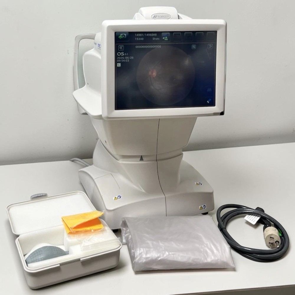
Need a comparable fundus camera available today? These units are in stock and ready to ship with a 90-day Digital Eye Center warranty.
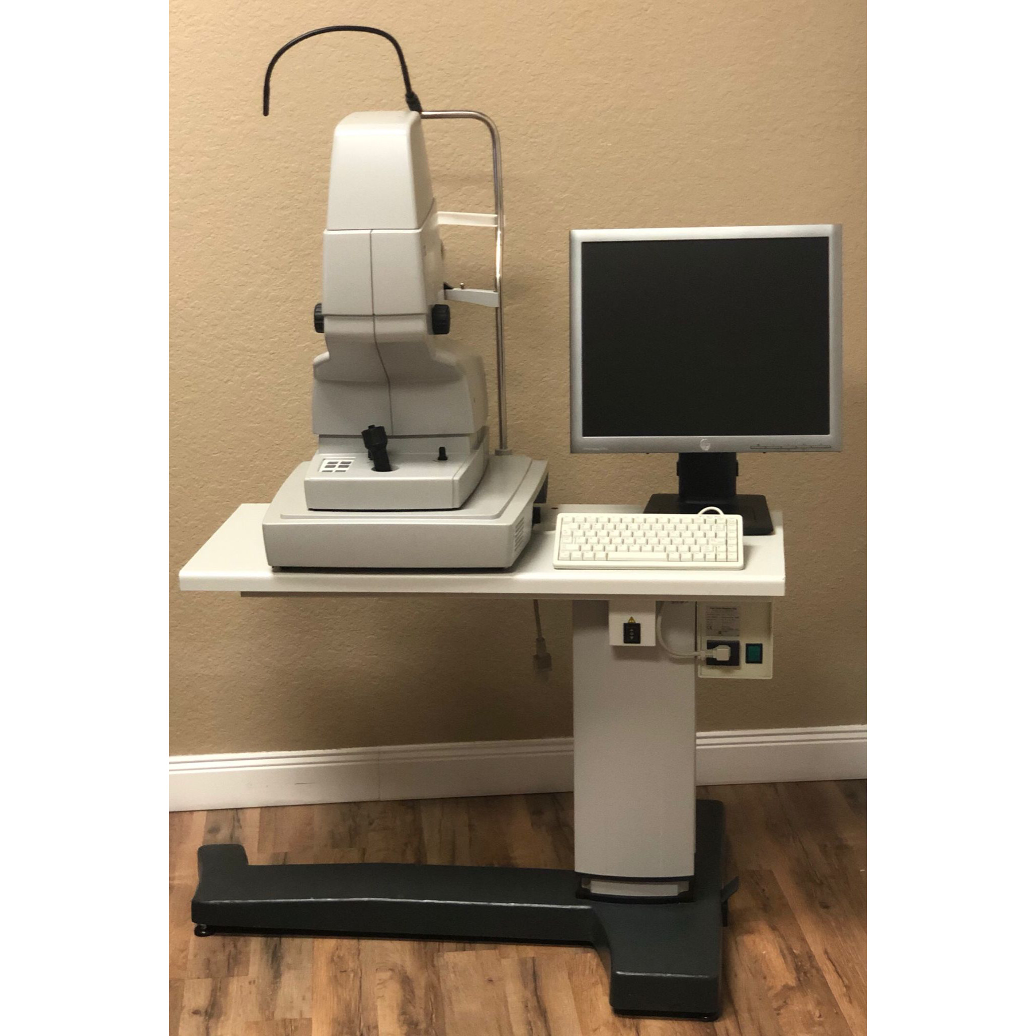
Prefer to wait for the Canon CR-2 Plus FAF Non-Mydriatic Retinal Camera- Used? Request availability and we’ll source one for you.
What’s Included
- Canon CR-2 Plus FAF retinal camera unit
- Fundus autofluorescence (FAF) imaging module
- Power supply and cables
- Original Canon documentation
- 90-day Digital Eye Center warranty
Condition & Warranty
Used. This Canon CR-2 Plus FAF has been inspected and verified for full non-mydriatic retinal imaging function — including color fundus, FAF acquisition, IR alignment, and autofocus — prior to sale. Standard cosmetic wear consistent with clinical use; full optical and functional integrity confirmed. Covered by a 90-day warranty on parts and workmanship.
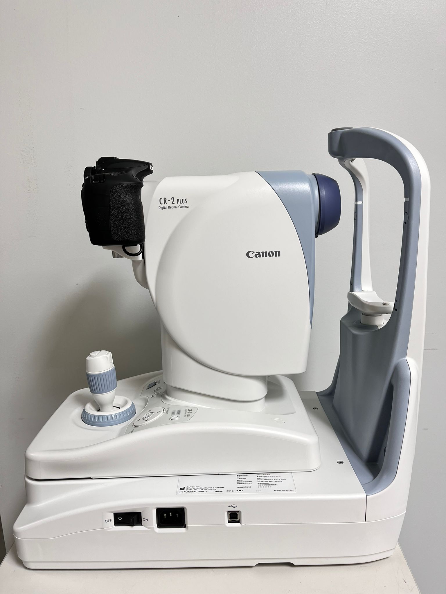
Non-Mydriatic Retinal Camera Canon CR-2 Plus FAF — Used Fundus Camera with Autofluorescence
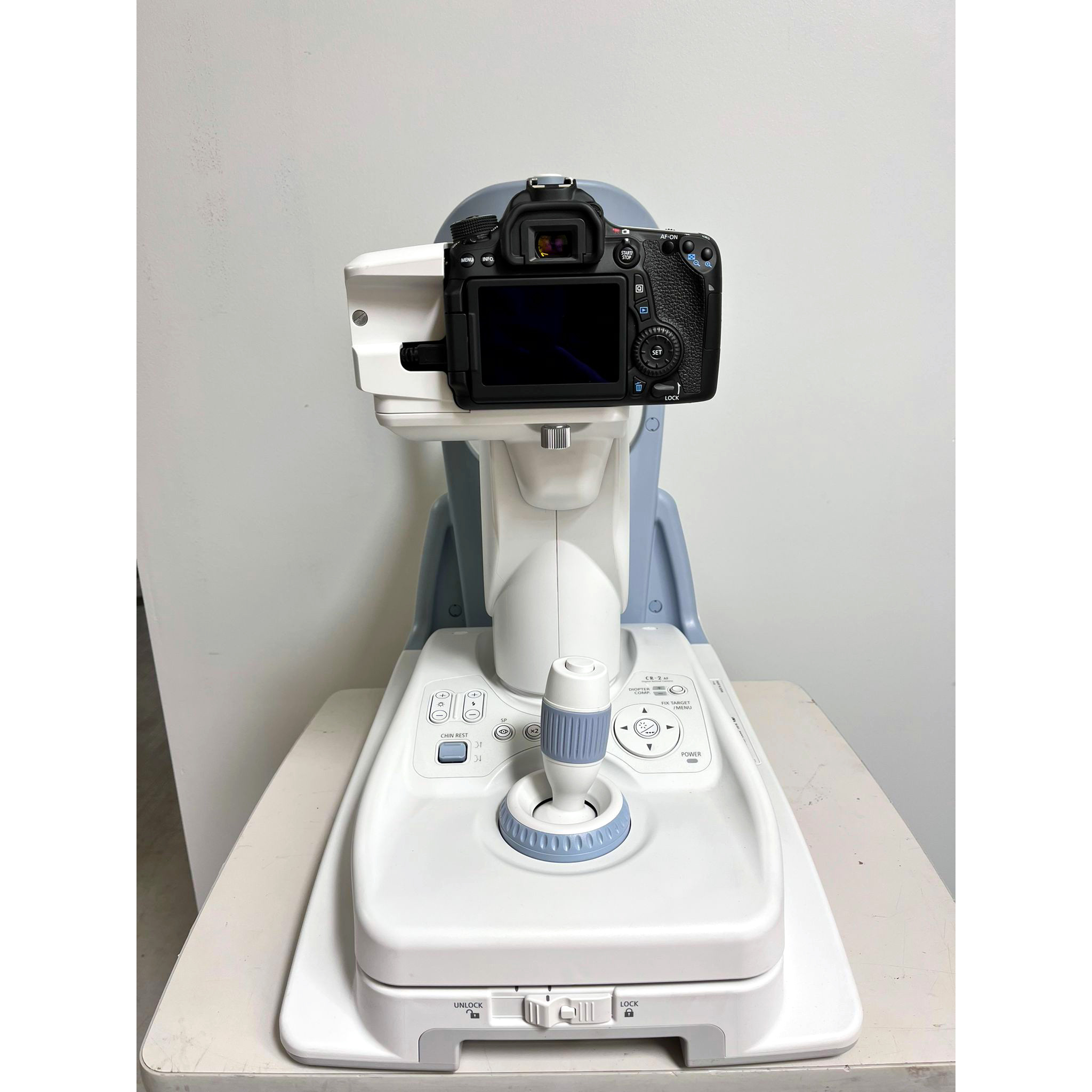 The Canon CR-2 Plus FAF is a professional tabletop non-mydriatic retinal camera combining color fundus photography, fundus autofluorescence (FAF), and infrared (IR) retinal imaging in a single non-dilated workflow. With a 45° field of view, a 4.0mm minimum pupil requirement (3.3mm in small-pupil mode), and ±18D diopter compensation, it handles the full spectrum of patient presentations — from routine posterior pole documentation to FAF-based evaluation of age-related macular degeneration, geographic atrophy, and inherited retinal dystrophies. Resolution of 63 lp/mm at center and 40 lp/mm at the periphery delivers image quality sufficient for clinical diagnosis and telemedicine grading. This used CR-2 Plus FAF is sold inspected and backed by Digital Eye Center’s 90-day warranty.
According to the National Library of Medicine, fundus autofluorescence imaging provides essential structural information about retinal pigment epithelium health in age-related macular degeneration — confirming FAF-capable non-mydriatic retinal cameras as critical tools for AMD monitoring, geographic atrophy tracking, and inherited retinal disease evaluation in clinical practice.
Why Practices Choose the Canon CR-2 Plus FAF Non-Mydriatic Retinal Camera
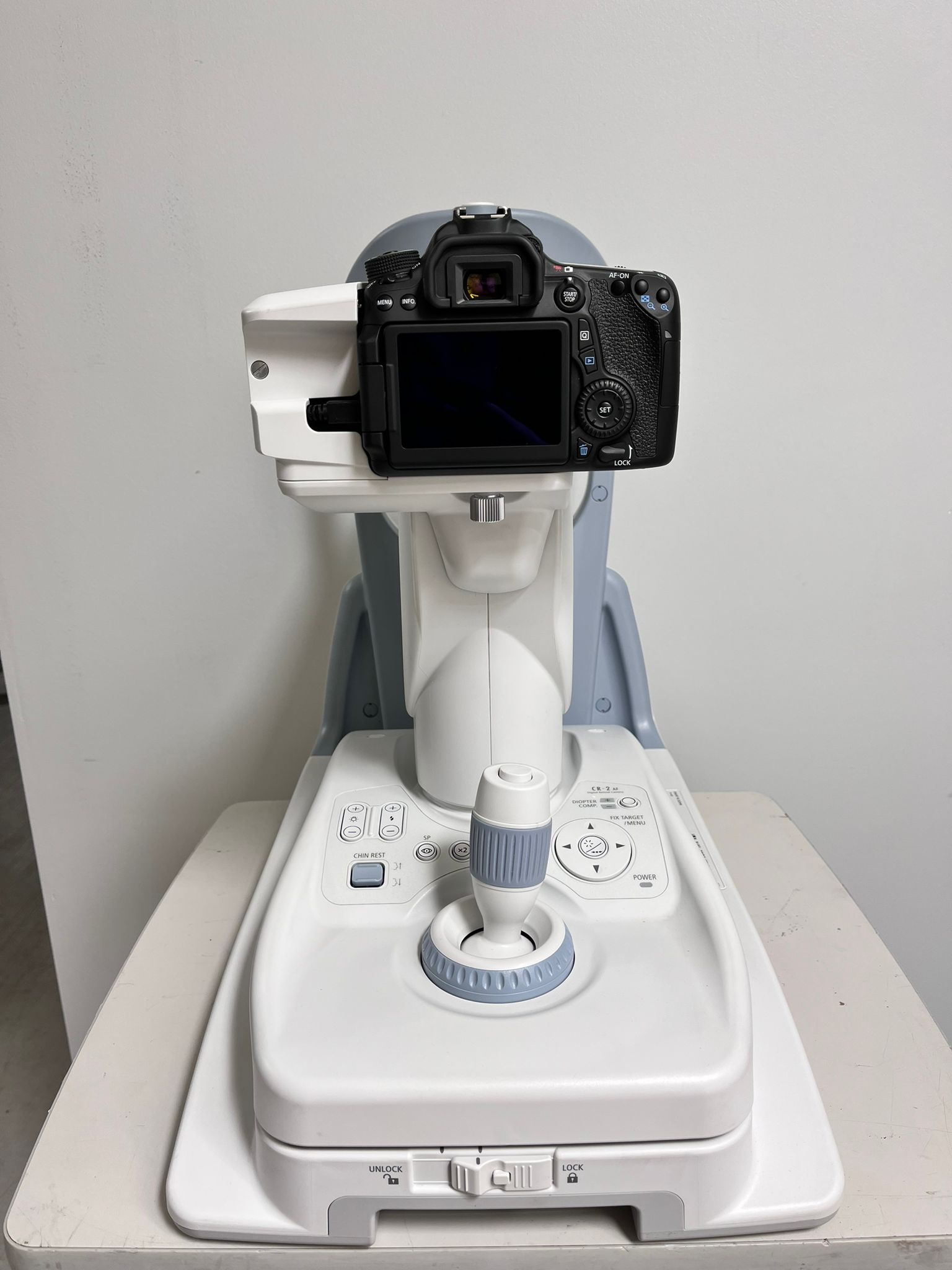 The CR-2 Plus FAF adds fundus autofluorescence to the proven Canon non-mydriatic platform — giving practices a single instrument for routine color photography and FAF-based retinal disease evaluation without a separate dedicated FAF camera.
- Fundus autofluorescence (FAF) imaging — single-button FAF acquisition visualizes retinal pigment epithelium lipofuscin distribution, detecting RPE atrophy patterns, geographic atrophy boundaries, and inherited dystrophy signatures before they appear on standard color photography.
- 45° non-mydriatic field of view — covers the posterior pole from optic disc to peripheral macula in a single frame without dilation, enabling rapid retinal documentation in routine exam lanes without a dilating agent or recovery period.
- 4.0mm pupil requirement (3.3mm small-pupil mode) — the small-pupil mode extends non-mydriatic imaging capability to elderly patients, post-surgical eyes, and patients on miotic medications, broadening the population that can be imaged without dilation.
- Color, FAF, and IR imaging modes — a single instrument delivers three complementary imaging modalities: color fundus for standard documentation, FAF for RPE and dystrophy assessment, and infrared for media clarity evaluation and patient alignment guidance.
- ±18D diopter compensation range — electric focus with a wide diopter range accommodates highly myopic and hyperopic patients, aphakic eyes, and post-surgical refractive changes without manual lens swapping.
- 63 lp/mm center resolution — image resolution sufficient for grading diabetic retinopathy features, drusen morphology, and disc margin detail in both color and FAF acquisition modes.
- Real-time IR alignment and preview — infrared aiming beam provides continuous alignment feedback during patient positioning, reducing acquisition attempts and improving image yield per patient encounter.
- Compact tabletop footprint — the CR-2 Plus mounts on a standard exam table, sharing lane space with other instruments and supporting multi-purpose exam room configurations.
Clinical Applications
- AMD and geographic atrophy monitoring — FAF maps RPE lipofuscin accumulation and atrophy boundaries for AMD staging, geographic atrophy progression monitoring, and treatment response documentation in anti-VEGF and emerging atrophy-targeted therapies.
- Inherited retinal disease evaluation — FAF patterns characterize Stargardt disease, Best vitelliform macular dystrophy, pattern dystrophies, and retinitis pigmentosa phenotypes, supporting genetic workup triage and longitudinal disease tracking.
- Diabetic retinopathy documentation — color retinal photography documents DR grade, provides retinal image for teleophthalmology DR grading networks, and supports diabetic eye exam compliance reporting for population health programs.
- Non-dilated screening programs — 4mm pupil threshold and small-pupil mode enable systematic non-dilated retinal imaging in primary care DR screening clinics, endocrinology offices, and community eye health programs.
- Pre- and post-surgical retinal documentation — color and FAF baseline photographs before vitreoretinal, cataract, or macular surgery support informed consent and provide post-operative comparison imaging for outcome documentation.
- Glaucoma disc documentation — color optic disc photographs with standardized illumination support glaucoma suspect baseline documentation and serial disc change assessment over time.
Who This Is For
- Comprehensive and medical ophthalmology practices needing a single non-mydriatic camera for color fundus documentation and FAF-based AMD and retinal dystrophy evaluation at a used price point.
- Retinal and macula specialists adding a non-dilated imaging workflow for routine monitoring visits — AMD follow-up, geographic atrophy tracking, and IRD surveillance — without occupying mydriatic imaging slots.
- Optometry practices performing non-dilated posterior segment screening and referring fundus photography reports, including FAF documentation for AMD co-management with retinal specialists.
- Diabetic retinopathy screening programs in primary care, endocrinology, and community health settings where non-dilated color fundus imaging drives DR detection without specialist referral for photography.
Technical Specifications
| Specification | Detail |
| Camera type | Tabletop non-mydriatic with FAF (color/FAF/IR) |
| Field of view | 45° |
| Minimum pupil diameter | 4.0mm standard; 3.3mm in small-pupil mode |
| Imaging modes | Color fundus, Fundus Autofluorescence (FAF), Infrared (IR) |
| Resolution (center) | 63 lp/mm |
| Resolution (periphery) | 40 lp/mm |
| Diopter compensation | ±18D (electric, automatic/manual) |
| Working distance | 35mm |
| Power | 100–240 VAC, 50/60 Hz |
| Condition | Used |
| Warranty | 90-day Digital Eye Center warranty |
Compare Non-Mydriatic Retinal Cameras
| Product | Type | Condition | Price |
| Canon CR-2 Plus FAF | Non-mydriatic, 45°, color + FAF + IR | Used | $4,700 |
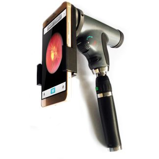
| Eyerobo iFlash Retinal Camera | Portable retinal camera — new | New | $9,025 |
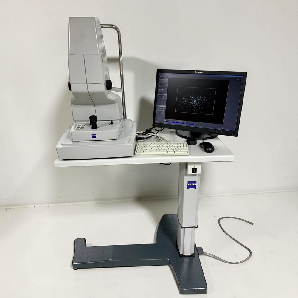
| Zeiss Visucam PRO NMFA | Tabletop non-mydriatic with FAF (color/FAF/IR) | Refurbished | $15,200 |
Need a newer portable option? See the Eyerobo iFlash portable retinal camera.
Questions about the Canon CR-2 Plus FAF? Contact us for availability and shipping details.