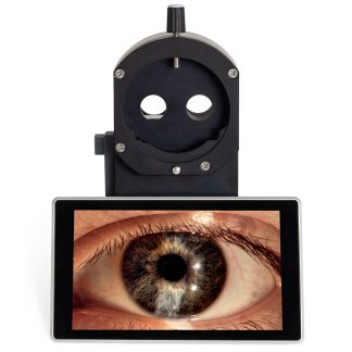
Description
The Optovue RTVue is an ultra-high speed, high resolution optical coherence tomography (OCT) retina scanner used for retina imaging and analysis.
It is based on the next generation Fourier-Domain Optical Coherence technology just emerging from clinical research in the last two years.
The ultra-high speed and high resolution features enable the FD-OCT to visualize the retinal tissue with ultra-high clarity in a fraction of seconds. myopic patients.
Tracking is available now for the Optovue RTVue SD-OCT.
Comprehensive Glaucoma Analysis with 3 in 1 registered scan
- Complete Disk/Rim/Cup Analylsis
- RNFL Map
- RNFL thickness at 3.45 mm diameter
Disk/Rim/Cup & RNFL thickness map
- AIG Study scan protocol
- Evaluate disk and cup shape and symmetry
- Calculate all stereo-metrics parameters e.g. rim volume, cup volume, C/D, etc.
- RNFL thickness map
- ~20,000 data points in 0.35 seconds

Retinal Thickness Map – 7 mm x 7 mm
- Evaluation of ganglion cell loss with full retina or inner retina thickness map
- Asymmetry assessment of superior and inferior hemispheres from thickness differential map – full or inner retina

RNFL thickness at 3.45mm diameter around disk
- Progressive and asymmetry analysis
- High repeatability with average data from 4 consecutive high density scans

Examine Retina with multiple Views
Multiple map views of Pathology:
- Full retinal & Inner retinal thickness maps
- ILM & RPE elevation maps
- Visualize fine detail beneath the map
- Quantify the height and size of lesions
- High quality near IR fundus image allows visualization of sub-surface lesions

Unprecedented image resolution and clarity in two perspective views registered on fundus image

Visualize Pathology in 3D

Quantitative progressive comparison after treatment

Visualize pathology in all perspective views
3D Scan in 2 seconds
B-Scan
- Control of perspective views
- Synchronized views of B-Scan, SLO location , 3D presentation
3D Interactive Presentation
- Rotate
- Fly-through B-Scans
- Zoom
- Position

Total Corneal Power
The Total Corneal Power Upgrade is the first FDA cleared SD-OCT based cornea power measurements which directly measures both the anterior and posterior curvatures rather than assuming an anterior/posterior relationship.
This methodology, coupled with 5 micron resolution SD-OCT technology, allows evaluation of post refractive patients with increased confidence.
Corneal powers from Optovue RTVue are entered into a unique online formula for IOL calculations.
Tracking
The Tracking feature for RTVue® provides exquisitely detailed B-scans of up to 16mm length. Capture high quality scans even on patients with fixation and movement issues as well as high myopic patients. Tracking is available now for the RTVue SD-OCT.
Specifications of the Optovue RTVue 100 OCT
| RTVue Scanner | OCT Image: 26,000 A-scan/second Frame Rate: 256 to 4096 A-scan/Frame Depth Resolution (in tissue) : 5.0µm Transverse Resolution: 15µm Scan Range: Depth: 2- 2.3mm Transverse: 2mm to 12mm Scan Beam Wavelength: λ=830±10nm, Exposure Power at pupil: 750µW |
| Fundus Imager | FOV: 32 o (H) x 23 o (V) Minimum Pupil diameter: 3.0 mm Illumination: Near IR |
| Patient Interface: | Working Distance: 22mm Motorized Focus Range: -15D to +12D |
| Computer Unit: | CPU: 3GHz Intel Dual-Core Processor RAM: 2GB |
| Foot print: | 1010mm(W) x520mm(D) |
| Retina Analysis: | |
|---|---|
| Line Scan: | 1024 A-Scan/Frame |
| High Definition Line Scan: | 4096 A-Scan/frame |
| Cross Lines: | A pair of 1024 pixel horizontal/vertical lines |
| HD Cross Lines: | a pair of 4096 pixel H/V lines |
| MM5 (Macular Map 5×5 mm) | – Inner/Outer Retinal Thickness Map – ILM/RPE Elevation Map |
| 3D Scan (Disk & Macular) in 2 seconds | – Simultaneous 3D, B-Scan, C-Scan, enface images. |
| Glaucoma Analysis: | |
| NHM4 (Nerve Head Map 4 mm Diameter) | – RNFL Thickness Map – Nerve Head disk, rim, and cup map – Nerve Head stereometric parameters |
| MM7 (Macular Map 7 x 7 mm) | – Inner/Outer Retinal thickness Map – Superior/Inferior Difference Map |
| RNFL 3.45mm | – Classical 3.45mm diameter circular scan – Average over 4 consecutive scans done in 76 msec |
Further Information
OCT Optovue RtVue