Description
This exact unit sold recently — but we specialize in sourcing refurbished ophthalmic equipment. Typical turnaround is 2–4 weeks once we confirm availability with our partners. Request a free quote with no deposit required.
Request Availability → Free quote · No obligation · 90-day warranty when shipped
What’s Included
- Zeiss Visucam 524 fundus camera main unit
- Acquisition computer with Zeiss Forum / VISUCAM software
- Chinrest and head positioning assembly
- All standard cables and power supply
- 6-month Digital Eye Center warranty on parts and workmanship
Condition & Warranty
Refurbished. This Zeiss Visucam 524 has been inspected, cleaned, and function-tested across all imaging modes to confirm optics, sensor, auto-focus, and auto-alignment are performing to specification. Covered by a 6-month warranty on parts and workmanship from Digital Eye Center.
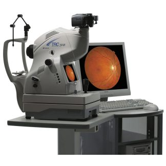
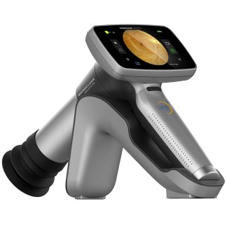
Zeiss Visucam 524 — Refurbished 24MP Non-Mydriatic Fundus Camera
 The Zeiss Visucam 524 is ZEISS’s flagship non-mydriatic fundus camera, combining a 24-megapixel sensor with proprietary ZEISS precision optics to deliver the highest-resolution color fundus photography available in a tabletop platform. This refurbished Zeiss Visucam 524 captures ultra-high-resolution detail across a 45° standard field with optional 30° zoom, producing images detailed enough to grade fine microaneurysms, neovascularization, and disc margin changes that lower-resolution cameras miss. The Zeiss Visucam 524 features auto-focus, auto-alignment, and non-mydriatic capability with a 3.6 mm minimum pupil diameter — keeping workflow moving in practices where dilation is impractical.
The National Library of Medicine (Sci Rep, 2025) confirms that high-resolution fundus photography enables the development of reliable automated diabetic retinopathy screening tools — underscoring why the image quality of the Zeiss Visucam 524 is critical for today’s AI-assisted clinical workflows.
Why Practices Choose the Zeiss Visucam 524
- 24-megapixel sensor — The highest-resolution sensor in the Visucam line delivers images with sufficient detail to detect early microaneurysms, fine neovascularization, and subtle disc changes that 12–16 MP cameras miss entirely
- ZEISS precision optics — Proprietary ZEISS lens system eliminates chromatic aberration and edge distortion, producing tack-sharp images from center to periphery across the full 45° field
- Non-mydriatic imaging (3.6 mm minimum pupil) — Captures diagnostic-quality images without dilation in the majority of patients, enabling faster clinic flow and broader population screening eligibility
- Multimodal imaging: color, red-free, FAF — A single platform covers color fundus photography for diabetic retinopathy grading, red-free imaging for RNFL assessment, and autofluorescence for AMD and macular dystrophy monitoring
- Auto-focus and auto-alignment — Automated acquisition minimizes the skill threshold for technicians and reduces the rate of ungradable images in high-volume diabetic screening workflows
- Zeiss Forum integration — Images are natively compatible with the Zeiss Forum imaging network, enabling multi-site sharing, longitudinal comparison, and integration with electronic health records
- Compact, robust design — Low-maintenance platform engineered for daily clinical use across ophthalmology, optometry, and endocrinology screening settings
Clinical Applications
 The Zeiss Visucam 524 sits at the top of the non-mydriatic fundus camera market for practices that require publication-quality retinal photography. Its 24 MP output is the standard used by retina subspecialists for disease progression documentation, legal-quality photography, and high-fidelity teleretinal programs where image quality determines whether remote grading is possible.
- Diabetic retinopathy grading (ETDRS-standard) — 24 MP resolution meets and exceeds the image quality standards required for Early Treatment Diabetic Retinopathy Study-level grading and telemedicine program submission
- AMD monitoring and documentation — High-resolution color and autofluorescence imaging tracks drusen load, geographic atrophy boundaries, and choroidal neovascularization over time
- Glaucoma optic disc photography — Resolves subtle cup-to-disc ratio changes, disc hemorrhages, and peripapillary RNFL defects that lower-resolution cameras cannot capture reliably
- Retinal vascular disease documentation — Branch and central retinal vein/artery occlusions, neovascularization, and laser treatment scar mapping
- AI-assisted screening integration — 24 MP image files meet the input specifications required by FDA-cleared diabetic retinopathy AI algorithms
- Macular disease and dystrophy surveillance — Red-free and FAF modes document photoreceptor and RPE health across the spectrum of inherited and acquired macular disease
Who This Is For
- Retina practices that need the highest available fundus image resolution for disease documentation, progression tracking, and treatment planning
- Ophthalmology groups running diabetic screening programs where image quality determines gradability and compliance with telemedicine payer requirements
- Academic medical centers and research programs requiring publication-quality retinal photography for clinical studies and AI dataset development
- Multi-specialty eye clinics seeking a single platform covering diabetic retinopathy, AMD, glaucoma disc photography, and macular dystrophy in one instrument
Technical Specifications
| Specification | Detail |
| Sensor Resolution | 24 megapixels |
| Standard Field of View | 45° |
| Optional Field | 30° (zoom) |
| Minimum Pupil Diameter | 3.6 mm (non-mydriatic) |
| Imaging Modes | True Color, Red-Free, Autofluorescence (FAF) |
| Optics | ZEISS precision optics |
| Acquisition | Auto-focus, auto-alignment |
| Software | Zeiss Forum / VISUCAM |
| Manufacturer | Carl Zeiss Meditec, Germany |
| Condition | Refurbished |
| Warranty | 6 months |
Compare Zeiss Visucam 524 with Other Visucam Models
| Feature | Zeiss Visucam 524 | Visucam Pro NM/FA (FAF/ICG) | Visucam Pro NM/FA |
| Sensor | 24 MP | 14 MP | 14 MP |
| Autofluorescence | FAF | FAF + ICG | FAF |
| Condition | Refurbished | Refurbished | Refurbished |
| Price | $22,300 | $17,100 | $15,200 |
Also see the Zeiss Visucam Pro NM/FA with FAF and ICG at $17,100, or the Zeiss Visucam Pro NM/FA at $15,200.
Similar Fundus Cameras In Stock Now
Need a comparable fundus camera available today? These units are in stock and ready to ship with a 90-day Digital Eye Center warranty.


Prefer to wait for the Zeiss Visucam – 524 – Fundus Camera – Refurbished? Request availability and we’ll source one for you.
Questions about the Zeiss Visucam 524? Contact us for availability and shipping details.