Description
What’s Included
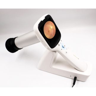
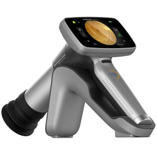
- iCare Eidon (Centervue) Ultra-Widefield Non-Mydriatic Retinal Camera
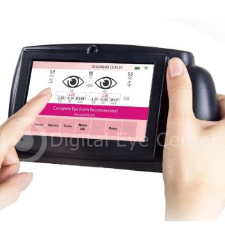
- Tablet controller
- Motorized electric table
- Chinrest and head positioning assembly
- All standard cables and power supply
- 90-day Digital Eye Center warranty on parts and workmanship
Condition & Warranty
Used. This iCare Eidon fundus camera has been inspected and function-tested to confirm all imaging modes, auto-alignment, and tablet control are operating correctly. Covered by a 90-day warranty on parts and workmanship through Digital Eye Center.
Eidon Fundus Camera — Used Ultra-Widefield Non-Mydriatic System
 The Eidon fundus camera (formerly Centervue Eidon, now iCare Eidon) is the benchmark ultra-widefield non-mydriatic fundus imaging system trusted by retina practices, diabetic screening programs, and optometry networks worldwide. This used Eidon fundus camera captures true-color confocal fundus images without dilation, covering a standard 60° field and extending to 100° with field-extension imaging — all in a single, compact tabletop platform. The Eidon fundus camera supports autofluorescence (FAF), infrared (IR), and red-free (RF) imaging modes on the same platform, and its confocal scanning laser architecture eliminates scattering artifacts common in flash-based cameras, delivering sharper detail in patients with media opacities, small pupils, or significant lens changes.
Research published in Scientific Reports (National Library of Medicine, 2022) confirms that ultra-widefield color fundus imaging significantly expands peripheral retinal visualization compared to standard-field cameras — detecting lesions that would otherwise be missed at 45° or 60° fields of view.
Why Practices Choose the Eidon Fundus Camera
- True-color confocal imaging — confocal scanning architecture eliminates scattering artifacts from flash illumination, producing sharper, higher-contrast images in small-pupil and media-opacity patients without dilation
- Ultra-widefield 100° field — standard 60° capture extends to 100° with field-extension mode, imaging the far peripheral retina where early diabetic lesions, lattice degeneration, and retinal tears most commonly appear
- Non-mydriatic acquisition — designed for undilated pupils as small as 3.3 mm, enabling efficient high-volume screening workflows without pharmacological dilation delays
- Multimodal imaging suite — true-color, autofluorescence, infrared, and red-free imaging modes available on the same platform, covering diabetic retinopathy grading, AMD monitoring, and choroidal nevus surveillance
- Auto-alignment and auto-capture — automated pupil detection and image acquisition reduces technician skill requirements and patient repositioning, improving throughput in high-volume diabetic screening environments
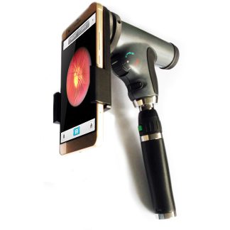
- Tablet-based control — intuitive touchscreen acquisition interface eliminates the need for a separate workstation; images transfer via network to the practice management system
- Compact footprint with electric table — built-in motorized table accommodates a wide range of patient heights and mobility needs, reducing setup time between patients
The iCare Eidon has become the preferred non-mydriatic camera for diabetic retinopathy telemedicine programs due to its image quality and peripheral coverage. Its integration with iCare’s cloud-based CLINIC software enables remote grading, AI-assisted analysis, and longitudinal comparison — workflows increasingly required by payers and ACOs for diabetic eye care quality metrics.
Clinical Applications
 The iCare Eidon’s 100° field extension and non-mydriatic confocal optics make it versatile across primary eye care, endocrinology, and retina subspecialty settings. In a standard diabetic screening program, a single technician can image both eyes of a patient — undilated — in under two minutes, with images immediately available for in-office or remote grading.
- Diabetic retinopathy screening and grading — ultra-widefield non-mydriatic imaging covers the peripheral retina where early NPDR lesions are most prevalent; peripheral NV and IRMA documented without dilation
- AMD staging and monitoring — true-color and autofluorescence modes document drusen load, geographic atrophy boundaries, and RPE changes for longitudinal comparison
- Peripheral retinal disease detection — 100° field extension captures lattice degeneration, retinal breaks, vitreoretinal traction, and peripheral neovascularization invisible to standard 45°–60° cameras
- Glaucomatous optic nerve documentation — high-resolution optic disc imaging with RNFL assessment supports early glaucoma detection alongside disc appearance monitoring
- Telemedicine and remote grading programs — image quality and peripheral coverage meet the requirements of diabetic teleretinal programs, enabling asynchronous review by remote graders
- Small-pupil and dense-media patients — confocal optics and sensitivity to 3.3 mm pupils allow imaging in cataract, vitreous floater, and non-dilating-pupil patients where flash cameras fail
Who This Is For
- Optometry practices adding retinal imaging for diabetic co-management, annual wellness exams, and insurance-reimbursed fundus photography
- Endocrinology and internal medicine groups running point-of-care diabetic retinopathy screening programs for their patient panels
- Ophthalmology offices seeking a high-throughput non-mydriatic camera for technician-driven diabetic and AMD screening before physician review
- Community health centers and mobile screening programs where efficient non-mydriatic throughput and compact size are critical
- Telemedicine retinal programs that require widefield fundus image quality for remote grading and AI-assisted diabetic retinopathy analysis
Technical Specifications
| Specification | Detail |
| Imaging Technology | Confocal scanning laser ophthalmoscope (non-mydriatic) |
| Standard Field of View | 60° |
| Ultra-Widefield (extension) | Up to 100° |
| Minimum Pupil Diameter | 3.3 mm (non-mydriatic) |
| Imaging Modes | True Color, Autofluorescence (FAF), Infrared (IR), Red-Free (RF) |
| Image Resolution | 14 MP |
| Acquisition | Auto-alignment and auto-capture |
| Control Interface | Integrated tablet |
| Patient Table | Motorized electric table (included) |
| Manufacturer | iCare (formerly Centervue), Italy |
| Condition | Used |
| Warranty | 90 days |
Compare Eidon Fundus Camera with Other Retinal Camera Options
| Feature | Eidon Fundus Camera | Zeiss Visucam Pro NM/FA (FAF/ICG) | Zeiss Visucam Pro NM/FA |
| Field of View | 60° / 100° widefield | 45° / 30° | 45° / 30° |
| Imaging Technology | Confocal scanning | Flash-based | Flash-based |
| Autofluorescence | Yes (FAF) | Yes (FAF + ICG) | Yes (FAF) |
| Condition | Used | Refurbished | Refurbished |
| Price | $26,000 | $17,100 | $15,200 |
Also consider the Zeiss Visucam Pro NM/FA with FAF/ICG for a conventional fundus camera with angiography capability, or the Zeiss Visucam Pro NM/FA for autofluorescence-only at a lower price.
Questions about the Eidon fundus camera? Contact us for availability and shipping details.