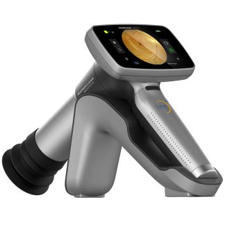
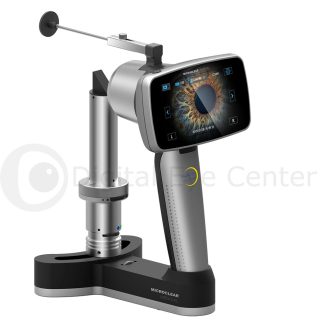
Description

Only Optos ultra-widefield technology can capture a 200° view (82%) of the retina in a single, high-resolution Optomap ® image in less than ½ second.
Optos Daytona offers autofluorescence imaging with green laser light and 3-in-1 Color Depth imaging ™. Unlike white light, low-powered laser wavelengths scan simultaneously, allowing a review of the retinal substructures in their individual laser separations:
- Color.
- Sensory (red-free).
- Choroidal.
Images are displayed in a consistent geometry that accurately represents anatomical features across the retina. Automatic image registration enables pixel-to-pixel comparisons of images across modalities and from visit to visit.
Features
- Non-mydriatic, non-contact imaging through 2 mm pupils and many cataracts.
- High image resolution shows fine detail across the retina (optic disc, macula, and periphery).
- Autofluorescence imaging with green laser light displays lipofuscin in the RpE.
- Eyesteering further extends the field of view past the vortex vessels in some cases.
- Stereo disc imaging.
- 3D Wrap ® for patient education.
- DiCOM compatible.
- Innovative software tools enhance image evaluation.
- Images are available immediately and stored electronically for future comparison or for use in telehealth applications.

Benefits
– Improves Practice Efficiency and Economics: Studies show that optomap images are faster to capture and easier to review than traditional patient examination techniques 1, 2.
Optomap enables practitioners to differentiate their practice, and an additional revenue stream can be generated.
– Enhances Clinical Decision-making: Early signs of many ocular pathologies and diseases may first present in the retinal periphery and can go undetected using conventional techniques and equipment.
More than 400 published and ongoing clinical trials, thousands of case studies, and testimonials show the long-term value of optomap imaging in the diagnosis, treatment planning, and patient engagement.
− Helps Prevent Vision Loss through Technological Innovation: optomap technology can image pathology past the vortex vessels, helping practitioners find disease sooner and manage it more effectively.
Technical Specifications
| General | |
| Trade Name | Daytona |
| Model Name | P200T |
| Imaging Modalities | Color Sensory (red-free) Choroidal Autofluorescence af |
| Resolution | optomap: 20 um optomap plus: 14 um |
| Wavelengths | Red Laser: 635 nm Green Laser: 532 nm |
| Exposure Time: | Less than 0.4 seconds |
| Footprint | Width: 440 mm/18 in Depth: 500 mm/20 in Height: 795 mm/32 in |
| Weight | 28 kg / 62 lbs |
| Laser Class | Laser safety class-1 following EN60825-1 and 21 CFR 1040.10 and 1040.11 |
| Communication Protocol | DICOM Compatible |