Description
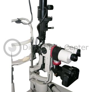
This Used Topcon OCT 3D Model 1000 includes:
- Topcon OCT 3D Model 1000
- PC with Pentium 3 or 4
- Original Table
- Printer
- 1 year Warranty
- On Site Setup and Training
Topcon OCT 3D Model 1000 Specifications
Advanced Features
- High Speed Scanning
- Wider Scanning Area
- Expanded Display of Depth
- Variety of Scanning Protocol
Scanning
- Spectral Domain OCT with TrueMap software provides incomparable details for clinical decision making
- Higher speed scanning to reduce artifacts due to eye movement
- A wider scanning area of 8.2 mm x 3 mm covers the macular and optic disc at once.
- The expanded display of 2.3 mm depth allows you to observe a large area of the optic disc or high-myopia.
- Fixation pattern, position are selectable and scanning center is movable in full scanning area. All these enable to quicklyl capture even the most difficult patient.
- Easy to use non-mydriatic retinal camera allows you to quickly compare the OCT image to the color fundus image.
- High myopia and cataract patients are easily captured with selectable fixation target
- An Auto-Z button that includes automatic Z lock button is now next to the omni control stick allowing you to quickly find the optimal scanning position.
TrueMap Software
The TrueMap software is intuitive and informative analysis tool of Topcon OCT 3D
With this proprietary software, unprecedented detailed imaging of the captured area allows you to see disease at its earliest stage.
Needless to say, early detection is critically important in Glaucoma. The Topcon OCT 3D Model 1000 with TrueMap software an unique display of 3D or 2D OCT image, color fundus image, RNFL topography image, and quantitative thickness map allows you to detect subtle variations of RNFL thickness, hopefully before the patient is symptomatic.
Data reproducibility
The incorporation of the non-mydriatic fundus images enables you to quickly see associate the OCT image with the color fundus image.
Topcon’s PinPoint Registration with TrueMap software enables easy and accurate chronological assessment of the same area of the retina greatly enhancing your ability to monitor the treatment of Glaucoma, AMD, CME and other eye diseases.
The Compare Function
The compare function allows you to compare side-by-side images and shows the differential map of thickness with PinPoint Registration and synchronization. This enables you to directly visualize the effectiveness of treatment.
PinPoint Registration
The TrueMap software alignment function quickly removes image artifacts due to axial eye movement while preserving the true anatomical structure of the retina, and displays 3D graphic.
This authentic unaveraged image will not miss even the slightest detail. Combined with PinPoint Registration the marked point can be reviewed anytime in the 3D scan, 2D Bscan, fundus image or in the thickness map.
Fundus image analysis
The captured color fundus image can be viewed RedFree, Blue only or at your preferable color balance with the RGB control function.
Cup-to-disc, line measurement, and area measurement tools are included to enhance your analysis of the image.
Tips for 3D image viewing
Layer segmentation, peeling, moving and cropping are useful tools for observing the 3D image in greater detail.
Mosaic Function
The panoramic fundus image and thickness map for RNLF or RPE covers the whole area of the fundus.
Quantitative Indication
The caliper function works in any mode of the B Scan image, thus the size of lesion can be measured at any angle.