Description
What’s Included
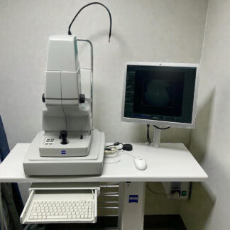
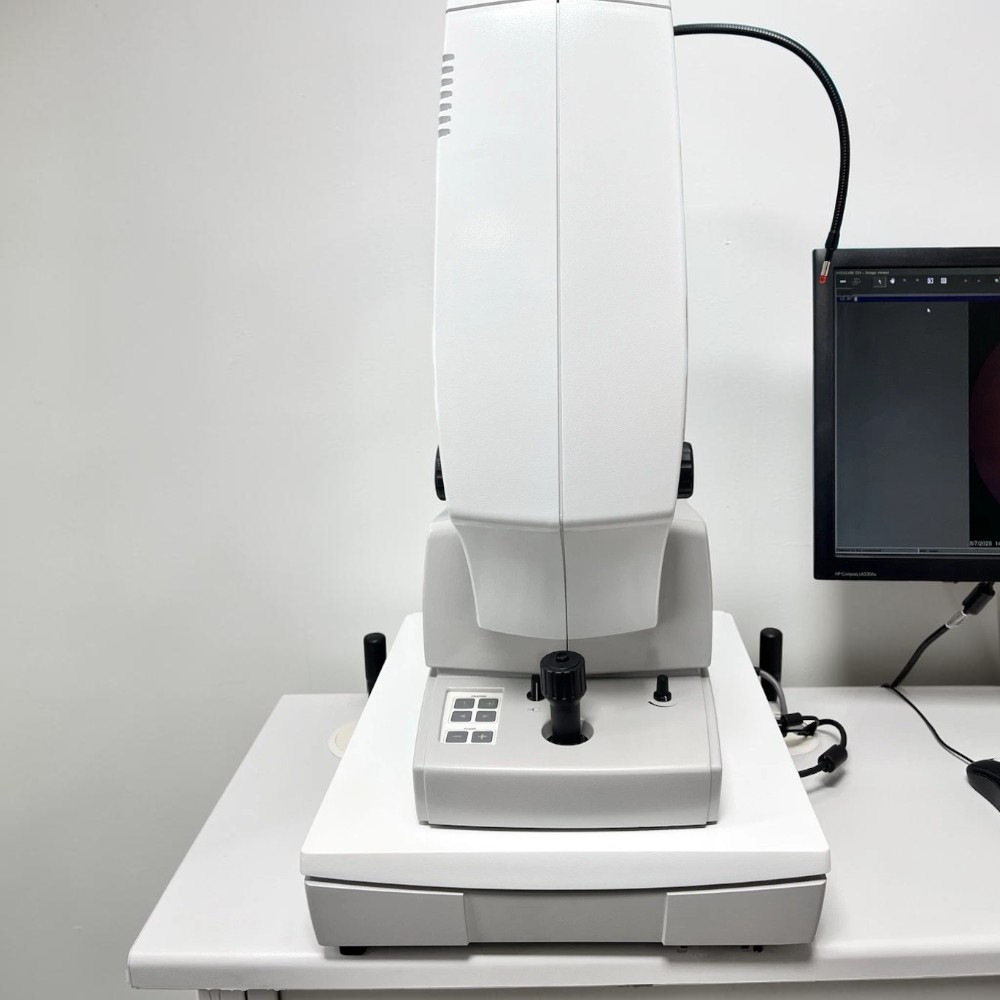
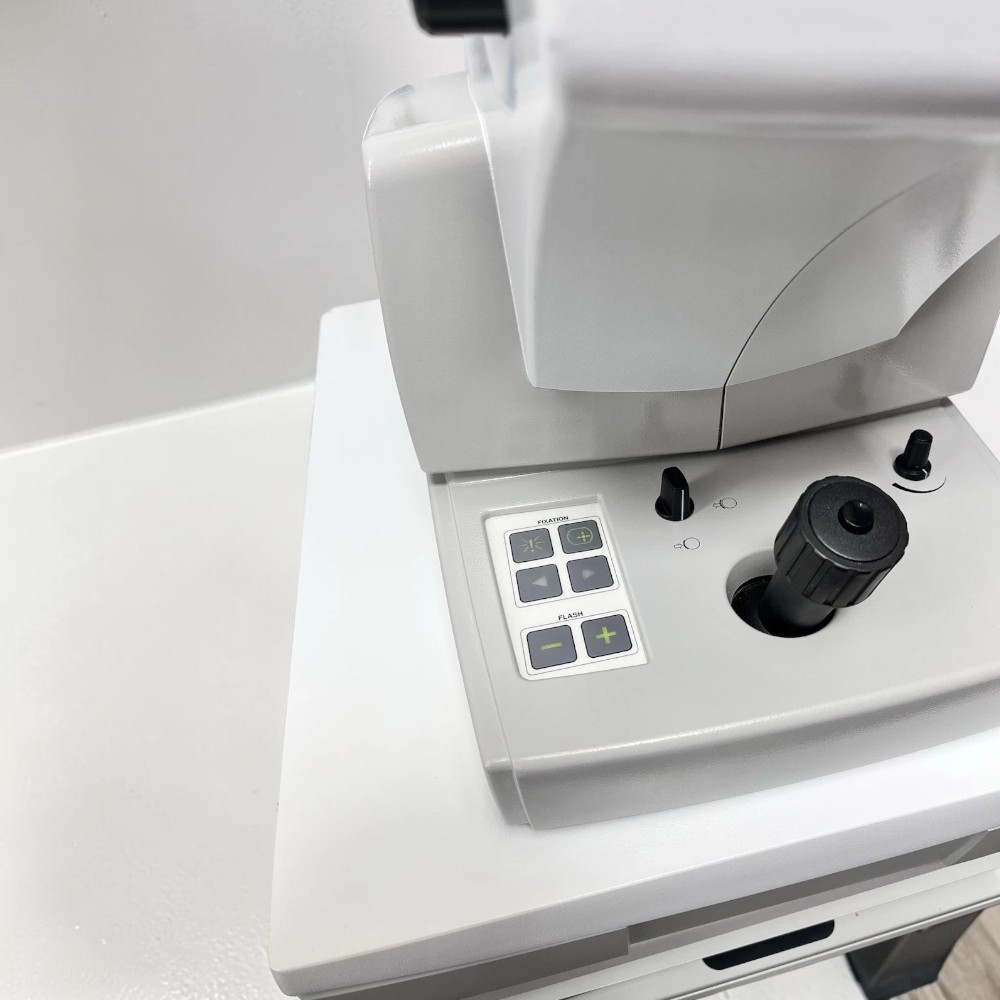
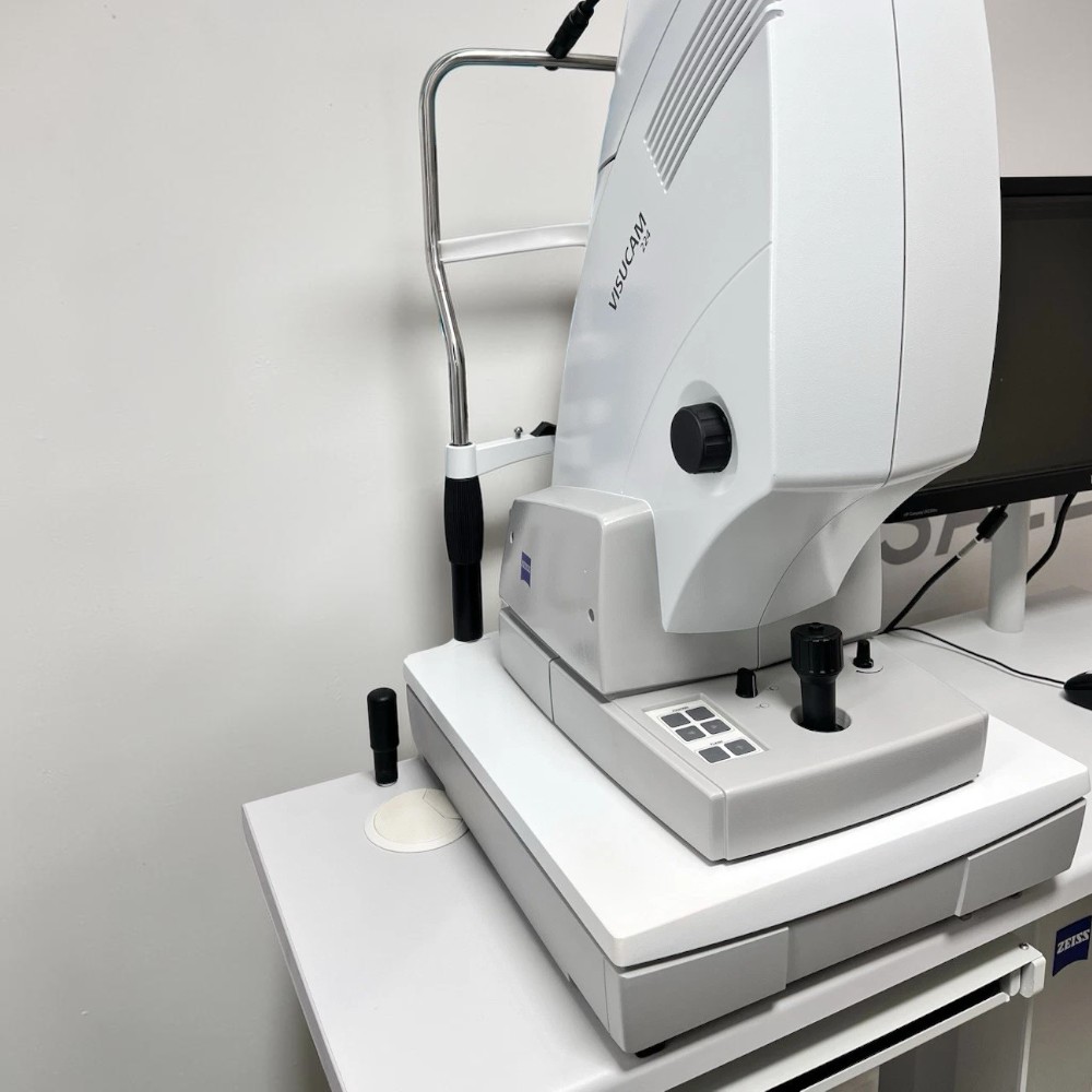
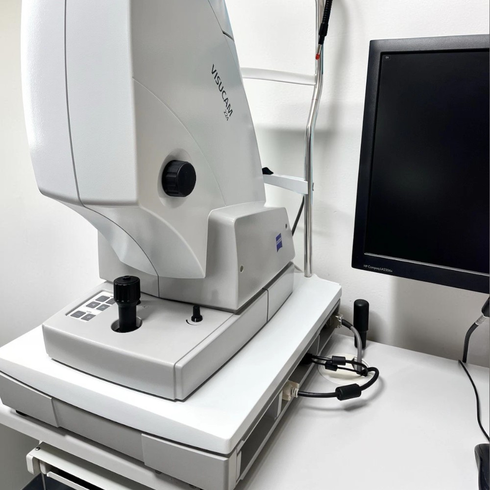
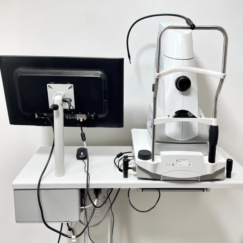
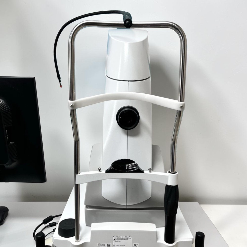
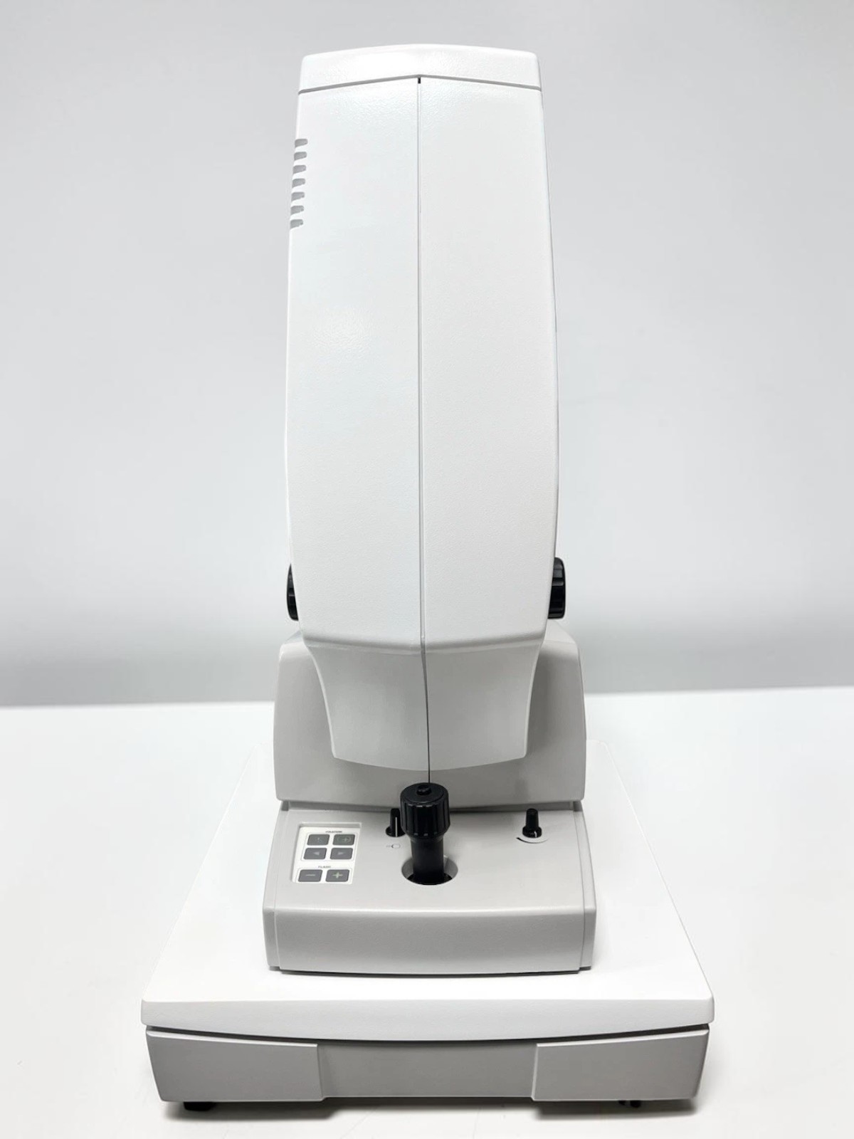
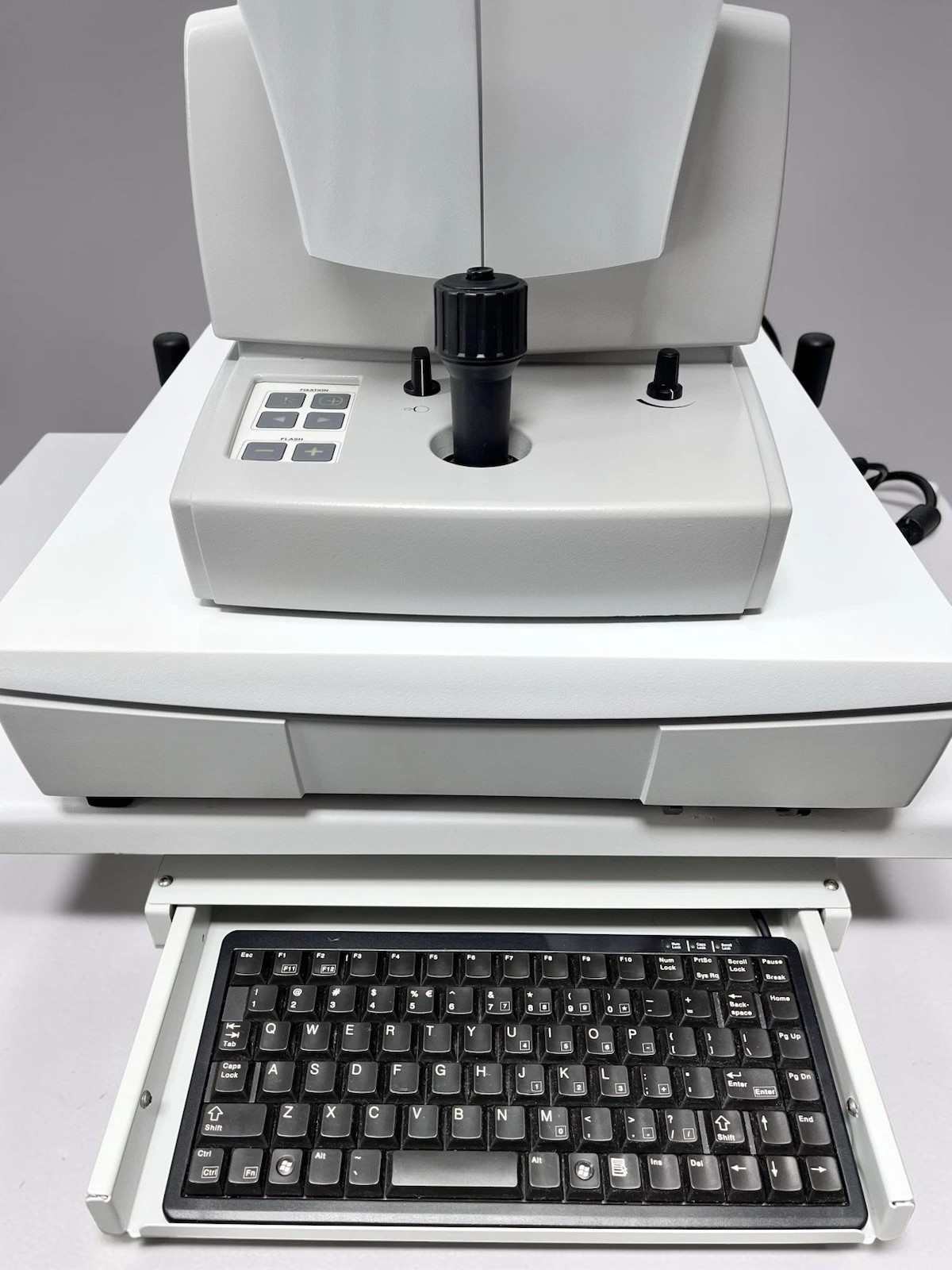
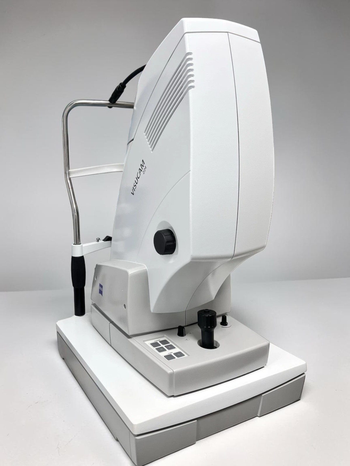
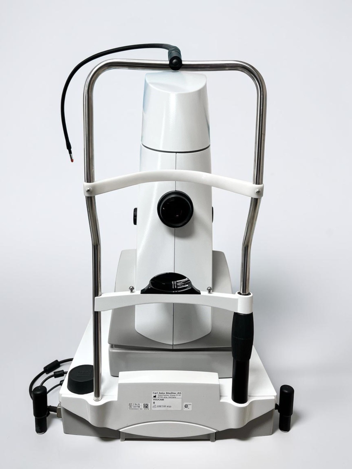
- Zeiss Visucam 224 Fundus Camera Main Unit
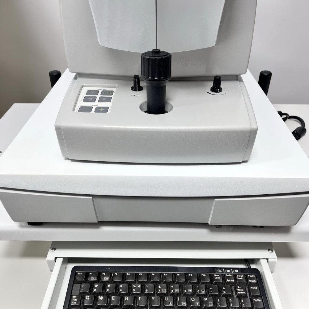
- Medical-Grade Electric Elevation Table
- Power cable
- Keyboard and mouse
- 90-day Digital Eye Center warranty
Condition & Warranty
Used. This Zeiss Visucam 224 package has been tested and is in excellent working order. It has been cleaned and is ready to use. The unit shows some signs of cosmetic use. Covered by a 90-day warranty on parts and workmanship.
Zeiss Visucam 224 — Used Non-Mydriatic Fundus Camera with Electric Table
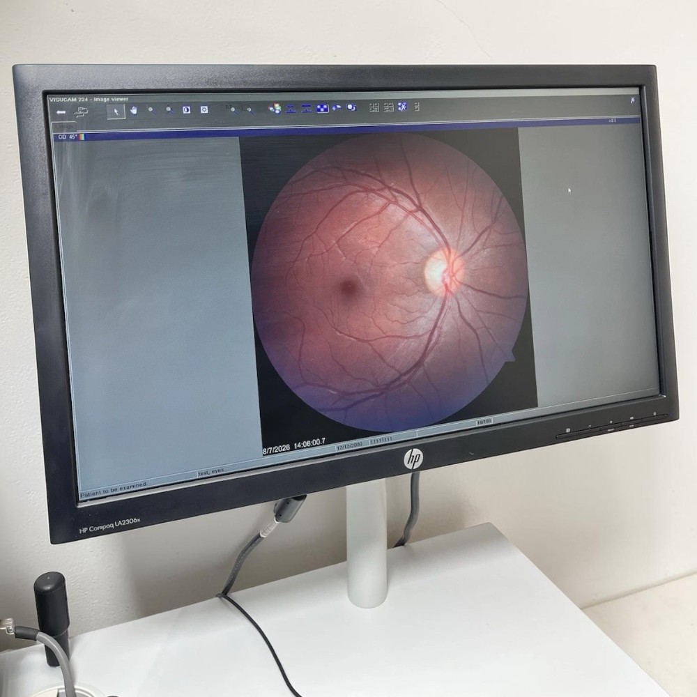
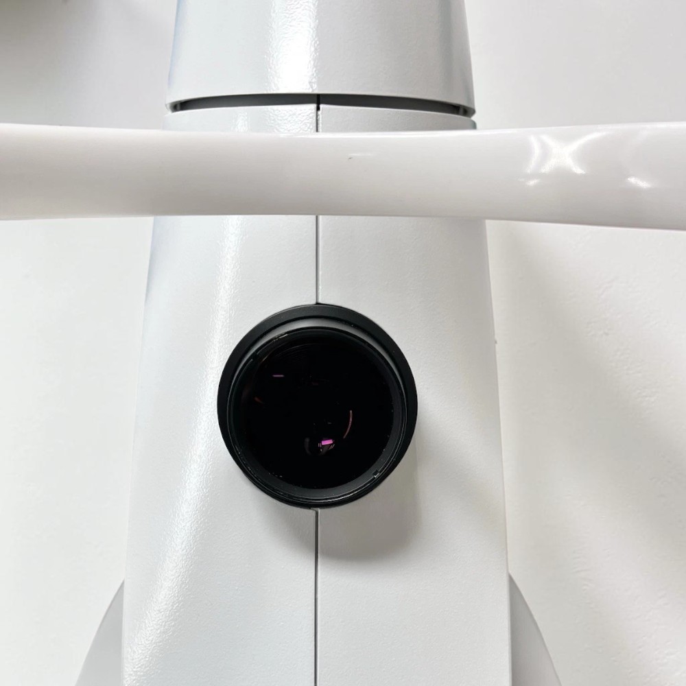
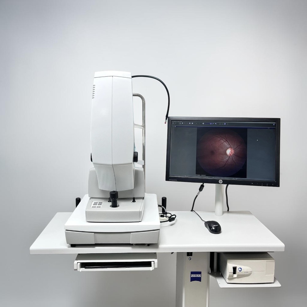 The Zeiss Visucam 224 is an elite, all-in-one digital retinal imaging system that pairs exceptional diagnostic versatility with a streamlined clinical workflow. Built around a high-resolution 24-megapixel optical sensor and renowned Zeiss glass optics, the Zeiss Visucam 224 produces color-accurate posterior segment images through pupils as small as 3.3 mm — without requiring diagnostic dilation drops.
This comprehensive workstation offers multiple clinical imaging modes, including True Color, Red-Free, Blue, and Red. Complete with a heavy-duty electric elevation table, this used Visucam 224 is a cornerstone asset for progressive eyecare practices.
A study published in the National Library of Medicine found that photographic non-mydriatic fundus screening markedly improves patient compliance with recommended eye examinations, supporting accurate baseline documentation for diabetic retinopathy, glaucoma optic nerve head evaluation, and macular pathology.
Why Practices Choose the Zeiss Visucam 224
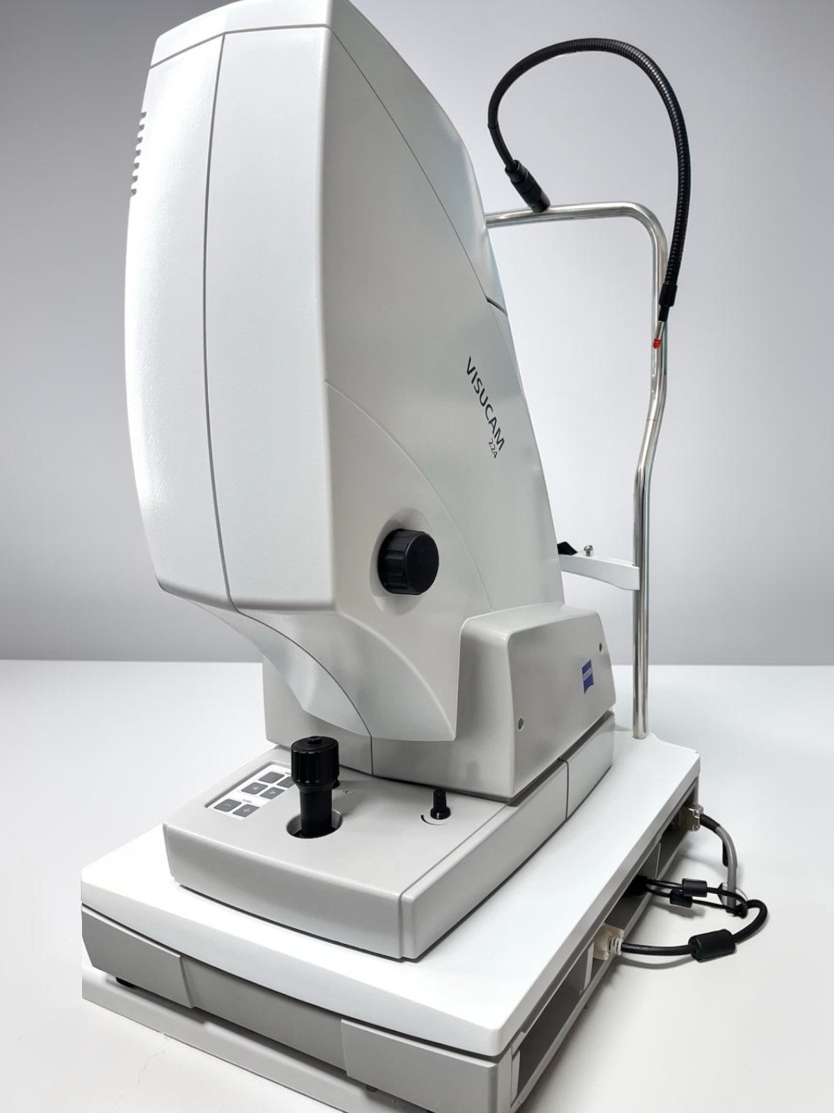
- High-Resolution 24 MP Sensor — Captures highly detailed retinal images, allowing clear digital zoom into subtle vascular and nerve fiber layer structures.
- Non-Mydriatic Small-Pupil Mode — Reliable captures through natural pupils down to 3.3 mm, improving patient comfort and exam-lane speed.
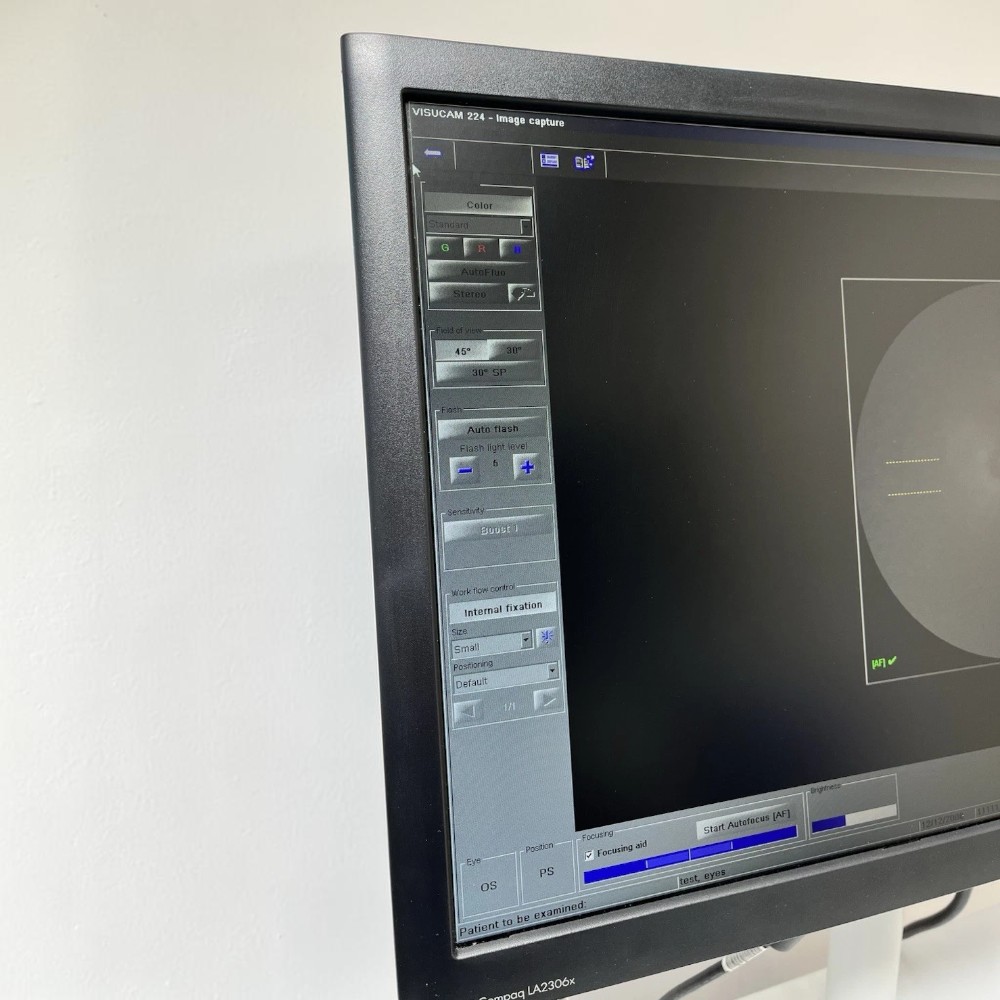
- Multi-Modal Diagnostic Versatility — True Color, Red-Free, Blue, Red, and External/Anterior Segment imaging.
- Auto-Focus and Auto-Flash Assistance — Automated exposure and alignment routines ensure consistent, high-quality images regardless of technician experience.
- Stereo Image Pair Capability — Captures overlapping optic disc images to support 3D spatial evaluation of cup-to-disc depth and rim geometry.
- Included Electric Elevation Table — Heavy-duty automated table provides fast, effortless height adjustments for patients of all sizes and mobility levels.
- Network & EMR Readiness — Compatible with ZEISS FORUM, DICOM, and standard EMR server networks for seamless electronic chart integration.
See the Zeiss Visucam 224 in Action
This official ZEISS overview walks through the VISUCAM 224/524 imaging platform and its non-mydriatic capture workflow.

Clinical Applications
- Diabetic Retinopathy Screening — Map microaneurysms, hemorrhages, and exudates across standard fields or multi-image montages.
- Glaucoma Optic Nerve Documentation — Analyze cup-to-disc ratios, neuroretinal rim tissue, and localized nerve fiber layer defects using high-contrast red-free filters.
- Macular Degeneration Progression Tracking — Document drusen, pigmentary changes, and geographic atrophy with color and red-free imaging over time.
- Anterior Segment and External Photography — Document corneal opacities, pterygia, and iris lesions via dedicated external imaging modes.
Who This Is For
- Comprehensive Optometrists and Ophthalmologists seeking a premier, multi-modal non-mydriatic camera to expand their diagnostic imaging capabilities.
- High-Volume Pre-Testing Suites that need fast, technician-friendly automated capture routines to maintain smooth patient flow.
- Retina and Glaucoma Clinics that require multi-spectral imaging modes (Red-Free, Blue, Red) paired with reliable digital archiving.
Technical Specifications
| Specification | Detail |
| Model | Zeiss Visucam 224 |
| Type | All-in-One Non-Mydriatic Fundus Camera |
| Field of View | 45° and 30° |
| Sensor Resolution | 24 Megapixels |
| Minimum Pupil Diameter | 3.3 mm (Small Pupil Mode) / 4.0 mm (Standard) |
| Imaging Modes | True Color, Red-Free, Blue, Red, Anterior Segment |
| Fixation Targets | Internal (programmable multi-point) & External |
| Table Type | Integrated Medical-Grade Electric Elevation Table |
| Condition | Used |
| Warranty | 90-day warranty on parts and workmanship |
Compare Non-Mydriatic Fundus Cameras
| Feature | Zeiss Visucam 224 | Topcon TRC-NW400 | Zeiss Visucam NM/FA |
| Sensor | 24 MP | Digital (auto-capture) | Integrated digital sensor |
| Field of View | 45° & 30° | 45° (30° via digital zoom) | 45° & 30° |
| Min. Pupil | 3.3 mm | 4.0 mm / 3.3 mm small-pupil | 3.3 mm |
| Fundus Autofluorescence (FAF) | Yes (standard) | No | No (premium variant only) |
| Fluorescein Angiography | No | No | Yes (standard) |
| Electric Table | Included | Not included | Included |
| Condition | Used | Used | Used |
| Price | $13,200 | $7,150 | $8,800 |
Also consider the Carl Zeiss Clarus 500 ultra-widefield fundus camera if your practice needs a wider capture area, or the more compact Topcon TRC-NW400 for entry-level screening.
Questions about the Zeiss Visucam 224? Contact us for availability and shipping details.