Description
This exact unit sold recently — but we specialize in sourcing refurbished ophthalmic equipment. Typical turnaround is 2–4 weeks once we confirm availability with our partners. Request a free quote with no deposit required.
Request Availability → Free quote · No obligation · 90-day warranty when shipped
Similar Fundus Cameras In Stock Now
Need a comparable fundus camera available today? These units are in stock and ready to ship with a 90-day Digital Eye Center warranty.
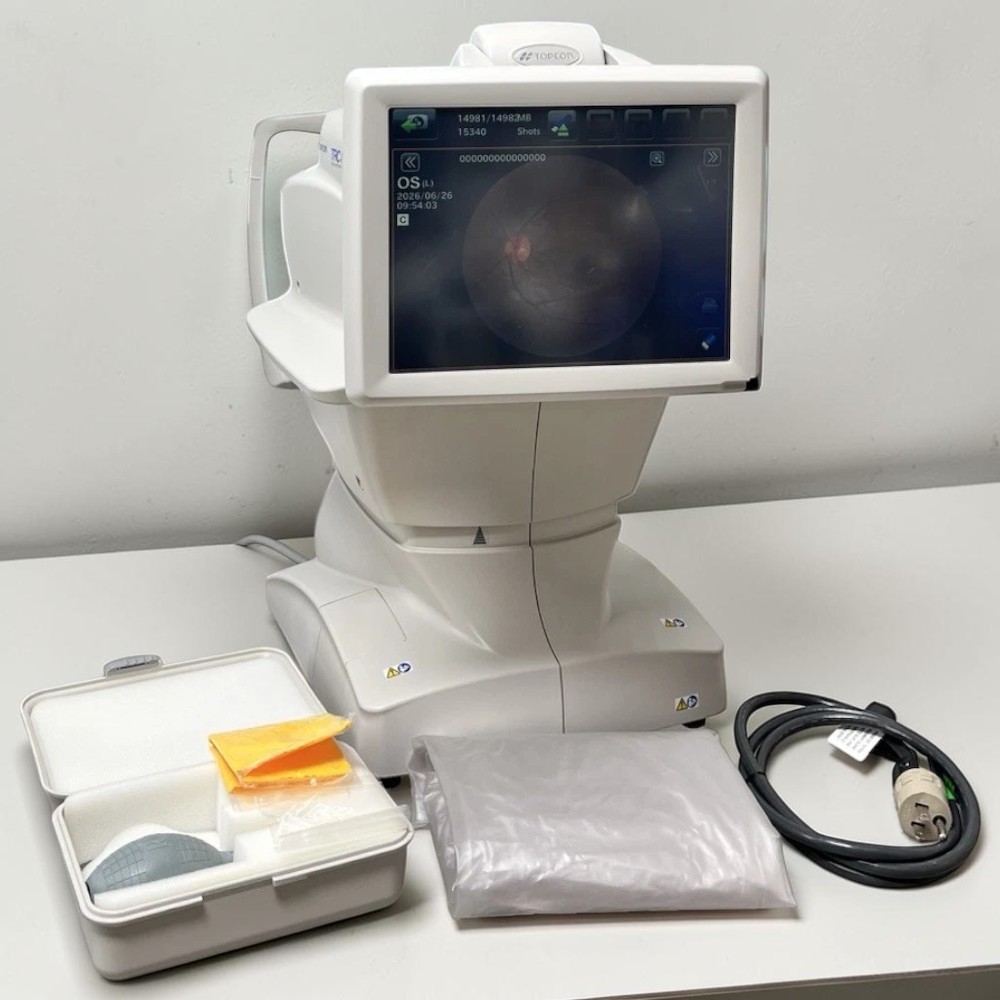
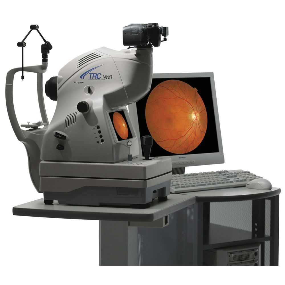
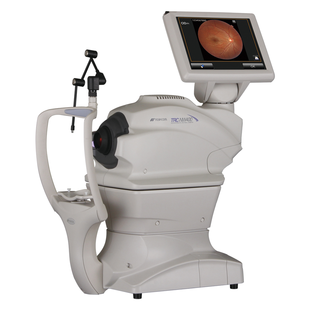
Prefer to wait for the Topcon NW6s NM Fundus Camera Used? Request availability and we’ll source one for you.
What’s Included
- Topcon TRC-NW6S non-mydriatic fundus camera
- Nikon 24.1 MP digital camera back
- PC with monitor
- Retinovue capture software
- Digital adapter
- Power cable
- Topcon NW6S user manual (PDF)
- 6-month Digital Eye Center warranty
Condition & Warranty
Used. This Topcon TRC-NW6S has been inspected and verified for full non-mydriatic retinal imaging function, digital camera capture, flash calibration, and alignment system operation prior to sale. Covered by a 6-month warranty on parts and workmanship.
Topcon Fundus Camera TRC-NW6S — Used Non-Mydriatic Retinal Camera with Nikon 24MP Back
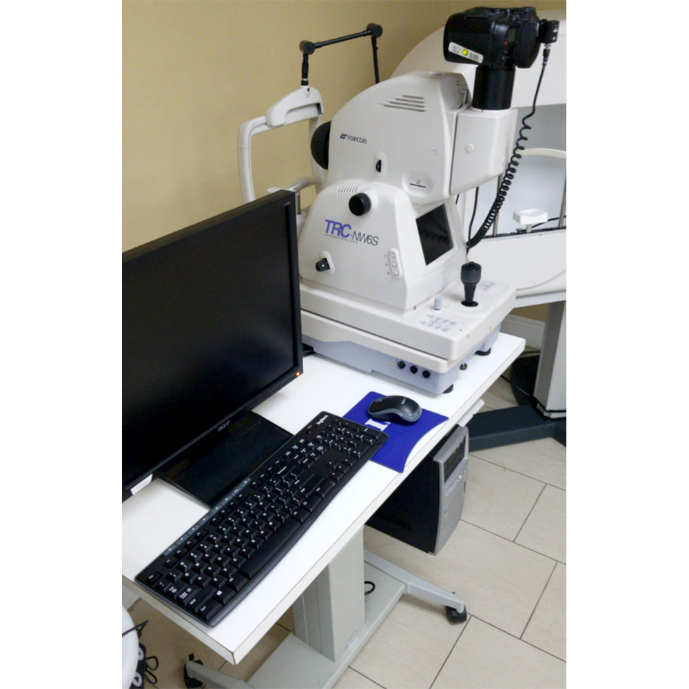 The Topcon fundus camera TRC-NW6S is a full-featured non-mydriatic tabletop retinal imaging system — capturing 45° and 30° field-of-view fundus photographs through pupils as small as 3.7mm, with no dilation required for most patients. This used Topcon fundus camera package includes the TRC-NW6S camera unit, a Nikon 24.1 megapixel digital camera back for high-resolution image capture, a dedicated PC and monitor, and Retinovue imaging software — providing a complete, ready-to-use retinal photography station. The dual-angle optical system, 8 peripheral fixation targets, and auto-adjusting 9-step flash intensity make the TRC-NW6S a reliable workhorse for daily diabetic retinopathy screening, optic disc documentation, and macular imaging. This Topcon fundus camera system is sold used, inspected, and backed by Digital Eye Center’s 6-month warranty.
According to the National Library of Medicine, non-mydriatic fundus cameras are clinically validated instruments for diabetic retinopathy screening programs — confirming that non-mydriatic capture delivers the image quality required for both ophthalmologist-led review and AI-assisted grading workflows.
Why Practices Choose the Topcon Fundus Camera TRC-NW6S
The TRC-NW6S delivers a complete non-mydriatic retinal photography workflow — dual-angle capture, automatic alignment, electric chinrest movement, and Nikon DSLR image quality — in a proven tabletop platform that integrates directly with IMAGEnet and Retinovue software. The combination of small pupil capability, panoramic peripheral imaging, and a high-resolution digital back makes it well suited for high-volume DR screening, optic disc documentation, and telemedicine image capture.
- Non-mydriatic capture through 3.7mm pupil — the 30° angle covers the macula and disc without dilation through a 3.7mm pupil; 45° coverage requires 4.0mm — enabling efficient, patient-friendly exams in the majority of clinical cases.
- Dual 45°/30° field of view — the optical system switches between 45° for a broad overview and 30° for higher-magnification macular and disc detail, with auto flash level adjustment when the angle is changed.
- Nikon 24.1 MP digital camera back — the included Nikon DSLR delivers significantly higher resolution than built-in CCD sensors, producing detailed retinal images suitable for grading, referral packages, and telemedicine programs.
- 8 peripheral fixation targets — internal fixation points guide systematic peripheral imaging in fixed, reproducible field positions for study protocols and longitudinal screening documentation.
- Auto flash intensity with 9-step manual override — flash level adjusts automatically when switching angles; manual 9-step control allows optimization for media opacity, pupil size, and image density.
- Panoramic mosaic imaging — optional mosaic module creates wide-field retinal overviews by stitching multiple peripheral fields, enabling comprehensive peripheral documentation from a single exam session.
- Split-line focus bars and two-spot alignment — alignment is confirmed by overlapping two bright spots on the monitor; focusing uses split-line bars visible in the operator view for fast, reliable image acquisition.
- Electric chinrest elevation — motorized chinrest buttons adjacent to the joystick enable quick patient height adjustment without interrupting the imaging workflow.
- IMAGEnet and Retinovue integration — R/L laterality, angle, and fixation position data output directly to IMAGEnet Lite; Retinovue software handles image management and workflow on the included PC.
Clinical Applications
- Diabetic retinopathy screening — non-mydriatic capture with 45° FOV enables single-field or two-field DR screening protocols without dilation, supporting high-volume primary care and endocrinology-based programs.
- Optic disc documentation and glaucoma follow-up — 30° field isolates the optic disc with higher magnification for cup-to-disc ratio documentation, hemorrhage detection, and longitudinal glaucoma monitoring.
- Macular imaging — 30° angle centered on the fovea captures age-related macular degeneration, diabetic macular edema, and macular pucker with the clinical detail required for referral and treatment decisions.
- Peripheral retinal documentation — 8 peripheral fixation targets and optional mosaic module support systematic retinal mapping for lattice degeneration, tears, and peripheral pathology documentation.
- Telemedicine retinal programs — Nikon 24MP capture produces high-resolution images for remote reading center grading, specialist referral, and AI-assisted diabetic retinopathy scoring platforms.
- Longitudinal retinal follow-up — reproducible fixation positions and consistent field documentation support side-by-side comparison of serial fundus photographs for progressive disease monitoring.
Who This Is For
- Ophthalmology and optometry practices running diabetic retinopathy screening or comprehensive retinal photography workflows that need a complete non-mydriatic fundus camera workstation at used pricing.
- Endocrinology and primary care offices implementing in-office DR screening programs with direct image management and telemedicine upload capability.
- Practices upgrading from older film-based or lower-resolution fundus cameras to a high-resolution Nikon DSLR digital back at a fraction of new equipment cost.
- Telemedicine and diabetic retinopathy reading center programs that require standardized, reproducible non-mydriatic fundus images for remote grading workflows.
Technical Specifications
| Specification | Detail |
| Imaging type | Non-mydriatic color fundus photography |
| Field of view | 45° and 30° (dual-angle) |
| Minimum pupil size | 3.7mm (30°); 4.0mm (45°) |
| Digital camera | Nikon 24.1 MP DSLR (included) |
| Fixation targets | 8 peripheral internal + central standard |
| Flash control | Auto-adjust (angle-linked); 9-step manual override |
| Diopter correction | -13D to +12D (standard); -12D to -33D (minus lens); +9D to +40D (plus lens) |
| Software | Retinovue (included); IMAGEnet Lite compatible |
| Workstation | PC with monitor included |
| Chinrest | Electric elevation (joystick-adjacent controls) |
| Power | AC 110/120/230/240V selectable; 50/60Hz |
| Weight (body) | 24.5 kg |
| Condition | Used |
| Warranty | 6-month Digital Eye Center warranty |
Compare Non-Mydriatic Fundus Cameras
| Product | Type | Condition | Price |
| Topcon TRC-NW6S with Nikon 24MP | NM Fundus Camera | Used | $5,100 |
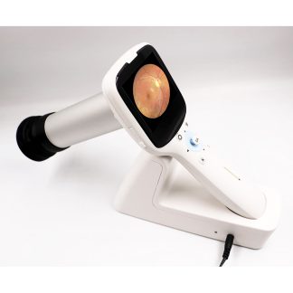
| Eyerobo iFlash Retinal Camera | Handheld Retinal Camera | New | $9,025 |
| Zeiss Visucam PRO NM/FA | NM Fundus Camera | Refurbished | $15,200 |
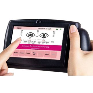
Looking for a handheld alternative? See the Eyerobo iFlash portable retinal camera.
Questions about this Topcon fundus camera? Contact us for availability and shipping details.