Description
What’s Included
- Optos California ultra-widefield fundus camera
- Tablet controller
- Multi-modality optomap® imaging software
- 90-day Digital Eye Center warranty on parts and workmanship
Condition & Warranty
Used. This Optos California fundus camera has been checked and certified by an authorized Optos technician to perfect cosmetic and working condition. Covered by a 90-day warranty on parts and workmanship from Digital Eye Center.
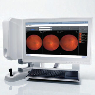
Optos California — Used Ultra-Widefield Fundus Camera
 The Optos California is the gold standard for ultra-widefield (UWF) retinal imaging, capturing 200° of the retina — approximately 82% of the total retinal surface — in a single sub-second acquisition without dilation in most patients. The Optos California delivers simultaneous multi-modality imaging: optomap® true-color, red-green, autofluorescence (green and blue), and fluorescein and ICG angiography from the same scan session. No other single-capture fundus device covers 200° of the retina — conventional cameras capture 45°–55°; even widefield systems cap at 100°–130°. This used unit has been checked and certified by an authorized Optos technician to perfect cosmetic and working condition.
The National Library of Medicine confirms that ultra-widefield imaging detects peripheral retinal lesions in diabetic retinopathy that are missed on conventional 45° fundus photography — the core clinical advantage the Optos California delivers over standard fundus cameras.
Why Practices Choose the Optos California
- 200° single-capture UWF imaging — approximately 82% of the retinal surface in one image; no stitching, no mosaic artifacts, no patient repositioning
- Multi-modality in one session — optomap® color, red-green, green AF, blue AF, fluorescein angiography, and ICG angiography from a single sitting without changing equipment
- Non-mydriatic through 2 mm pupil — cSLO laser technology captures high-quality images through small pupils and most cataracts; reduces or eliminates dilation for most patients
- Auto-montage to 220° — automated stitching combines images to visualize up to 97% of the retina in a single presentation view
- Peripheral lesion detection — identifies far-peripheral neovascularization, lattice degeneration, retinal breaks, and peripheral atrophy invisible on standard 45° imaging
- Sub-half-second capture — extremely fast acquisition reduces motion blur and patient discomfort, critical for elderly, pediatric, and low-cooperation patients
- Autofluorescence (green and blue) — RPE health mapping for AMD staging, geographic atrophy delineation, and CSCR evaluation without angiography dye
Clinical Applications
 The California’s 200° field makes it the dominant platform for population-level diabetic retinopathy screening and subspecialty retina practices where peripheral pathology drives management decisions. In a standard diabetic teleretinal screening program, the California’s single non-dilated image replaces the four to seven 45° images otherwise required to cover equivalent retinal territory — reducing capture time per patient and reading burden.
- Diabetic retinopathy screening — peripheral neovascularization, microaneurysms, and IRMA detected in a single non-dilated image; PDR staging with far-peripheral NV documentation
- AMD and geographic atrophy — blue autofluorescence maps RPE atrophy extent and pattern for AREDS2 staging and treatment planning
- Peripheral retinal disease — retinal breaks, lattice degeneration, retinoschisis, and peripheral degenerations requiring vitreoretinal referral
- Fluorescein angiography — full-field FA from disc to far periphery; leakage, non-perfusion, and neovascularization mapping across the entire posterior and mid-peripheral retina
- ICG angiography — choroidal vascular imaging for pachychoroid spectrum disease, polypoidal choroidal vasculopathy, and indeterminate macular lesions
- Uveitis and inflammatory disease — peripheral vasculitis, periphlebitis, and snowbank documentation invisible on posterior-pole-only imaging
- Telemedicine and remote reading — single optomap® image contains full retinal context; remote graders assess DR severity, referral urgency, and treatment response from one file
Who This Is For
- Diabetic retinopathy screening programs requiring non-dilated high-throughput imaging with single-capture coverage of the full retina
- Retina specialists needing far-peripheral documentation for vitreoretinal surgery planning, post-laser follow-up, and peripheral pathology referrals
- Comprehensive ophthalmology practices adding UWF imaging as a revenue and clinical differentiation tool alongside standard fundus photography
- Academic and research centers with UWF-based clinical trials requiring standardized 200° image sets
- Teleretinal reading programs where single-image retinal coverage is essential for efficient grader throughput
Technical Specifications
| Specification | Detail |
| Field of View | 200° single capture (82% of retina) |
| Auto-Montage | Up to 220° (97% of retina) |
| Imaging Modalities | optomap® color (rg/rgb), Red-free, Green AF, Blue AF, FA, ICG |
| Imaging Technology | Confocal scanning laser ophthalmoscope (cSLO) |
| Minimum Pupil Diameter | 2 mm (non-mydriatic in most patients) |
| Capture Speed | <0.5 seconds per image |
| Laser Wavelengths | 532 nm (green), 633 nm (red) |
| Condition | Used — certified by authorized Optos technician |
| Warranty | 90 days |
Compare Optos California with Other Fundus Camera Options
| Feature | Optos California | Optos California P200DT | Zeiss Visucam PRO NMFA |
| Field of View | 200° UWF | 200° UWF | 45° conventional |
| Autofluorescence | Green + Blue AF | Green + Blue AF | FAF included |
| FA / ICG | Both | Both | Both (NMFA model) |
| Non-mydriatic | 2 mm pupil | 2 mm pupil | Non-mydriatic |
| Price | $34,500 | $38,500 | $17,100 |
Also consider the Optos California P200DT for a newer model, or the Zeiss Visucam PRO NMFA for a conventional fundus camera with FA/ICG at a lower price.
Questions about the Optos California? Contact us for availability and shipping details.