Description
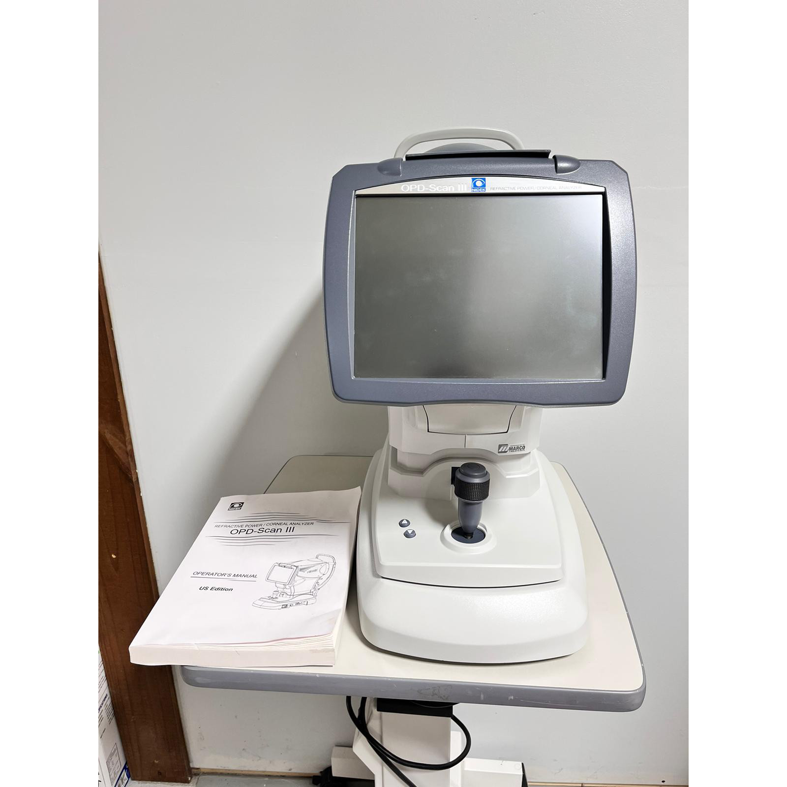
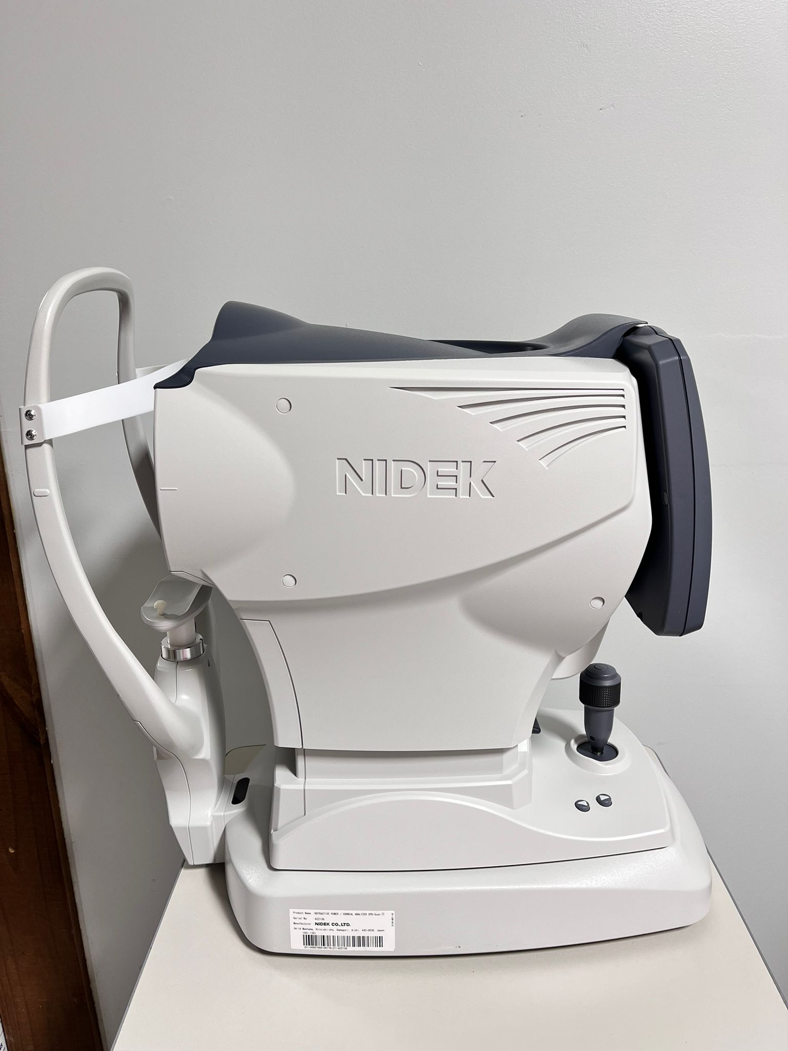
The Nidek OPD-Scan III is a comprehensive diagnostic tool offering various features and benefits to ophthalmologists. One of the most notable features is its ability to measure corneal topography, which helps identify irregularities on the surface of the cornea that can impact vision.
The device also offers a range of wavefront measurements, which help to identify aberrations in the eye and guide the selection of appropriate corrective lenses. The Nidek OPD Scan III also offers high-resolution imaging capabilities, allowing ophthalmologists to capture detailed eye images for diagnostic and documentation purposes.
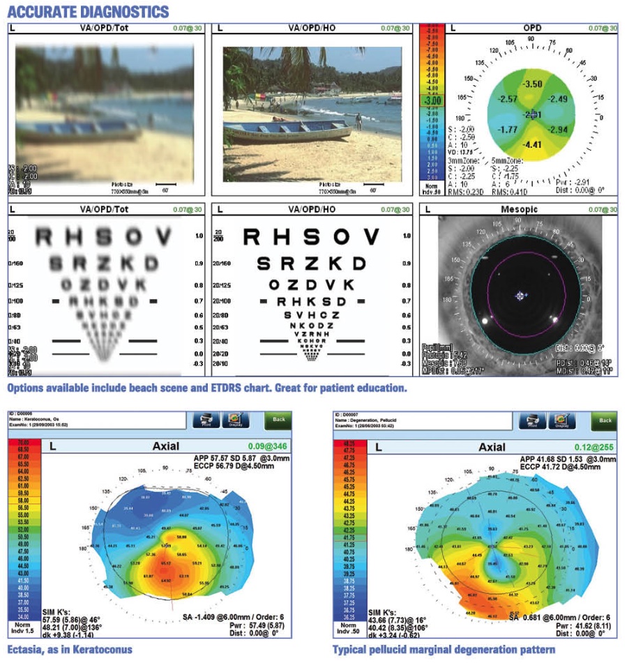
The device can also detect irregularities in the cornea that may impact vision, such as corneal ectasia, keratoconus, and irregular astigmatism. The Nidek OPD Scan III can also assess the eye’s overall health, including the lens and retina, and monitor changes over time.
Nidek OPD-Scan III Intelligent Technologies
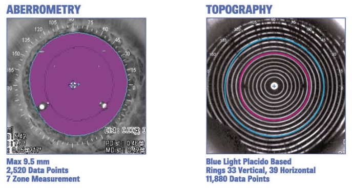
Over 20 diagnostic measurements are acquired in 10 seconds. Easy alignment and automatic data capture ensure accurate readings. Wavefront data is gathered from available zones up to a 9.5 mm area, adding the capability to calculate mesopic refractions. Blue light, 33-ring, and Placido disc topography are collected in one second.
Options to View Data include:
|
|
|
|
|
|
|
|
|
|
|
|
Nidek OPD-Scan III Unique Features
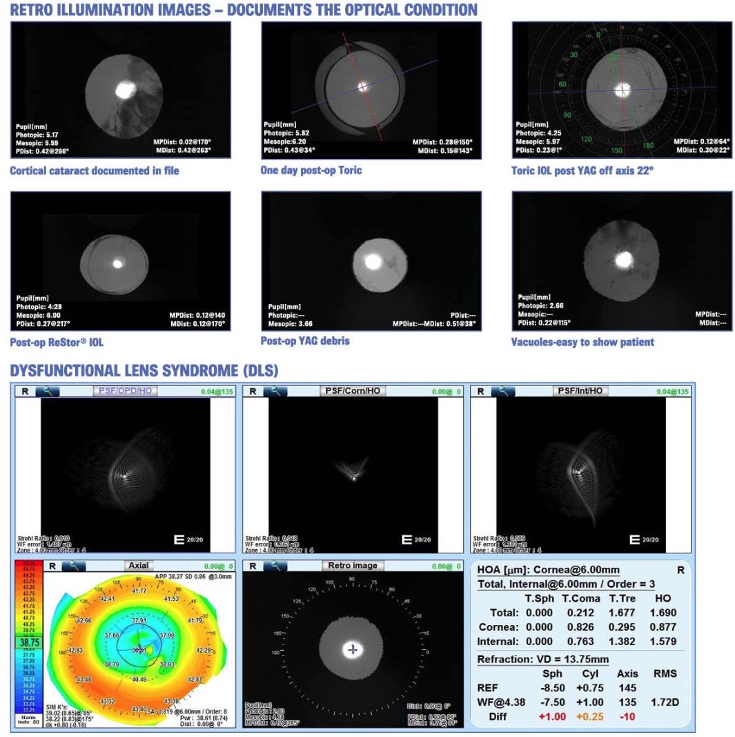
Cataract and refractive surgeries have now been taken to another dimension. Detailed patient summaries are available in seconds. Pre-op toric axis alignment can be mapped to the iris or other physical landmark positions. Retro illumination images can be used post-op to verify IOL axis alignment.
The above map measures a prior myopic LASIK patient with Dysfunctional Lens Syndrome (DLS). The Point Spread Function (PSF) maps show that the cornea contributes to the problem, but most of the patient’s issue is lenticular change. The patient thought she needed another LASIK treatment when the lens changed. A refractive lens exchange is recommended.
Nidek OPD-Scan III Day and Night Wavefront Refractions
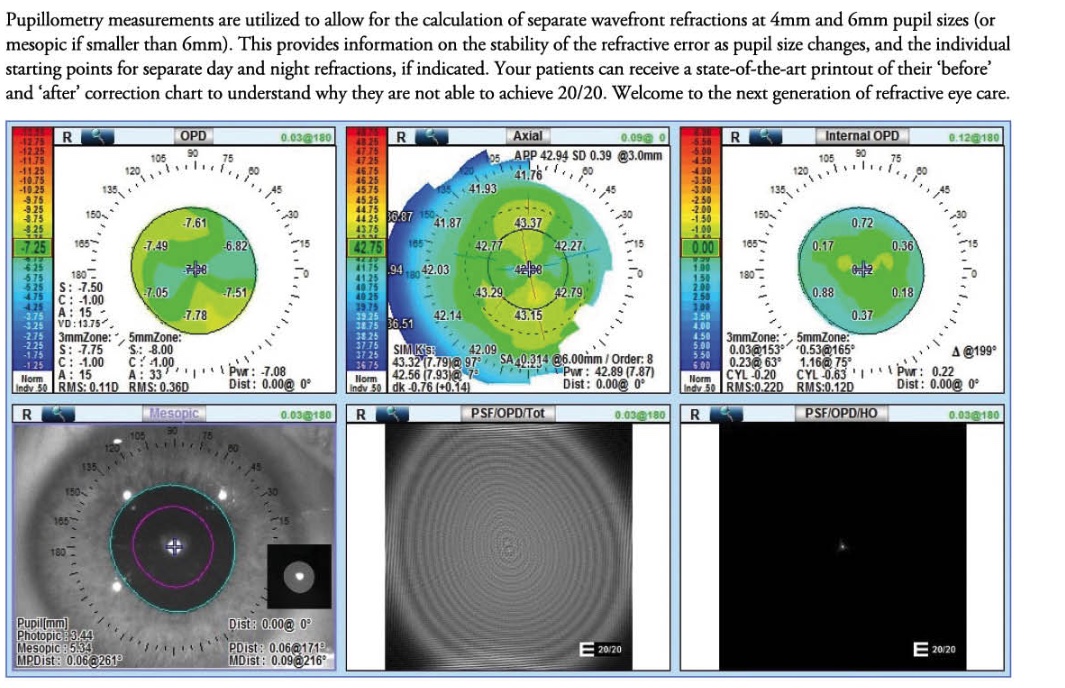
Pupillometry measurements are utilized to calculate separate wavefront refractions at 4mm and 6mm pupil sizes (or mesopic if smaller than 6mm). This provides information on the stability of the refractive error as pupil size changes and the individual starting points for separate day and night refractions if indicated. Your patients can receive a state-of-the-art printout of their “before” and “after” correction chart to understand why they cannot achieve 20/20. Welcome to the next generation of refractive eye care.
Available Data Displays include:
|
Captured in 10 seconds
|
Nidek OPD-Scan III: Multi-Modality Functions
The new OPD-Scan III is the latest diagnostic/refractive instrument that serves the practice as an autorefractor, keratometer, pupillometer, corneal topographer, and wavefront aberrometer.
Functional Highlights
- 10 seconds eye measurement
- Auto Alignment
- Auto tracking
- Auto capture
- Electronically adjustable chinrest
- Touch screen keyboard
- Verification after each measurement
- Easily integrates with EPIC and TRS-5100
IOL Applications
- APP – Average Pupil Power for Post Myopic LASIK calculations
- Angle Kappa, Angle Alpha
- Corneal Aberrations, including Corneal Coma and Spherical Aberration
- Pupillometry – photopic and mesopic pupils
- Corneal Topography
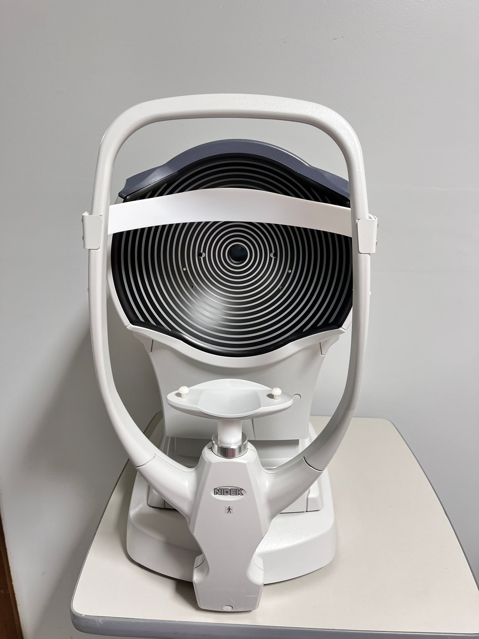
- Placido Rings for detection of any Ocular Surface Disease (OSD)
- Zernike Graph of Total, Corneal and Internal Aberrations
- White to White corneal diameter measurements
- Retro Illumination – Displays post-op Toric lens markings, opacities, etc.
- ECCp-Effective Central Corneal Power for IOL power calculation
- Toric IOL Summary to mark axis pre-op
- Eye image can allow for marking the cornea based on landmarks
- Cataract Summary displays the pertinent data together
- Point Spread Function graphs and VA simulation charts
OPD-Scan III Specifications
Power Mapping |
|
| Spherical power range | -20.00 to +22.00 D |
| Cylindrical power | 0.00 to +-12.00D |
| Axis | 0 to 180 deg. |
| Measurement area | 2.0 to 9.5 mm (7-zone measurement) |
| Data points | 2,520 points (7 x 360) |
| Measuring time | <10 seconds |
| Measurement method | Automated objective refractions (dynamic skiascopy) |
| Mapping methods | OPD, Internal OPD, Wavefront maps, Zernike graph, PSF, MTF graph |
Corneal Topography |
|
| Measurement rings | 33 vertical, 39 horizontal |
| Measurement area | 0.5 to 11.00 mm (r-7.9) |
| Dioptric range | 33.75 to 67.5 D |
| Axis range | 0 to 359 deg |
| Data points | More than 11,880 |
| Mapping methods | Axial, instantaneous, “Refractive”, Elevation, Wavefront maps, Zernike graph, PSF, MTF graph |
General Information |
|
| Working distance | 75 mm |
| Auto tracking | X-Y-Z directions |
| Observation area | 14 x 11 mm |
| Operating System | Windows 10 embedded |
| Display | 10.4-inch color LCD touch panel |
| Printer | Built-in thermal type line printer for data print
External color printer (optional) for map print |
| Power supply | 100 to 240 Vac
50/60 Hz |
| Power consumption | 110 VAC |
| Dimension /Mass | 286 (W)x525(D) x 530(H) mm / 23 kg |

