Description
What’s Included
- Heidelberg Spectralis OCT Mode 3 main unit
- Chinrest and forehead rest assembly
- Patient fixation target system
- Motorized patient table / instrument base
- Acquisition computer with Heidelberg Eye Explorer software
- All standard cables and power supply
- 6-month Digital Eye Center warranty on parts and workmanship
Condition & Warranty
Refurbished. This Heidelberg Spectralis OCT has been inspected, cleaned, and function-tested to confirm all imaging modes and TruTrack eye tracking are operating to specification. Covered by a 6-month warranty on parts and workmanship through Digital Eye Center.
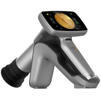
Heidelberg Spectralis OCT — Refurbished Spectral-Domain OCT
 The Heidelberg Spectralis OCT is the gold standard spectral-domain optical coherence tomography system used in leading retina and glaucoma practices worldwide. This refurbished Heidelberg Spectralis OCT Mode 3 delivers the same clinical precision as new — featuring 85,000 Hz scan speed, 3.9 µm axial resolution, and the proprietary TruTrack Active Eye Tracking technology that locks each scan to an exact retinal location with 1 µm AutoRescan reproducibility. No other OCT platform matches the Heidelberg Spectralis OCT for longitudinal disease monitoring. The Mode 3 also simultaneously captures a confocal scanning laser ophthalmoscope (cSLO) image alongside every OCT B-scan, enabling precise anatomical registration and true point-to-point comparisons at every follow-up visit.
Research published in Ophthalmology (National Library of Medicine) confirms that spectral-domain OCT reliably detects preperimetric glaucoma — structural damage that precedes any measurable visual field loss — making the Heidelberg Spectralis OCT an indispensable tool for early-stage disease detection.
Why Practices Choose the Heidelberg Spectralis OCT
- TruTrack Active Eye Tracking — compensates for involuntary eye movements in real time, ensuring every scan lands on the exact same retinal location regardless of patient fixation quality
- AutoRescan 1 µm reproducibility — follow-up scans are automatically registered to baseline, enabling detection of sub-clinical progression changes invisible on other platforms
- 85,000 Hz A-scan rate — ultra-fast acquisition reduces motion artifacts and enables high-density raster scanning across the entire posterior pole in seconds
- 3.9 µm axial resolution — resolves individual retinal layers including the ganglion cell complex, RNFL, and photoreceptor layer with unmatched clarity for structural analysis
- Simultaneous cSLO + OCT — confocal scanning laser image captured in parallel with every B-scan provides anatomical context and enables precise lesion localization
- BluePeak Autofluorescence (488 nm) — detects lipofuscin accumulation in the RPE, critical for monitoring geographic atrophy, Stargardt disease, and other macular dystrophies
- Modular upgrade platform — Mode 3 is field-upgradeable; practices can add MultiColor imaging, widefield, OCT-A, or anterior segment modules without replacing the base unit
- HEYEX 2 Software — unified platform for acquisition, analysis, and longitudinal trending with automated layer segmentation and normative database comparisons
The Spectralis Mode 3 is the most widely deployed configuration in academic medical centers and high-volume retina practices. Its optional panning camera headset and exterior touch panel allow technicians to make acquisition adjustments without disrupting the patient — a workflow advantage particularly appreciated in practices running multiple lanes.
Clinical Applications
 The Spectralis’s 1 µm AutoRescan reproducibility and simultaneous cSLO registration make it the reference platform for longitudinal retinal and glaucoma monitoring across all subspecialties. Point-to-point comparison across visits detects changes well below the threshold visible on competing systems, enabling earlier intervention in AMD, DME, and glaucoma progression.
- Glaucoma early detection and progression monitoring — RNFL and ganglion cell analysis with TruTrack registration catches structural progression before visual field changes are detectable on perimetry
- AMD staging and geographic atrophy monitoring — drusen volume, subretinal fluid, and RPE health tracked longitudinally with point-to-point reproducibility across months and years
- Diabetic macular edema quantification — central subfield thickness and total retinal volume measurements with automated layer segmentation guide anti-VEGF injection timing
- Retinal vascular disease imaging — high-resolution B-scans and cSLO fundus images document BRVO, CRVO, and macular ischemia with anatomical precision
- Macular dystrophy and hereditary retinal disease — BluePeak autofluorescence identifies RPE dysfunction patterns in Stargardt, Best disease, and cone dystrophies
- Neuro-ophthalmology — optic nerve head morphology, RNFL thinning patterns, and macular ganglion cell loss support diagnosis of optic neuritis, papilledema, and compressive optic neuropathy
Who This Is For
- Retina subspecialty practices seeking the highest longitudinal tracking precision for AMD, DME, and retinal vascular disease management
- Glaucoma specialists requiring structural OCT with TruTrack reproducibility to reliably detect progression in early or suspect glaucoma cases
- Academic medical centers and teaching hospitals that need a research-grade OCT platform with broad imaging module compatibility
- Multi-specialty eye clinics that see both anterior and posterior segment patients and plan to leverage the Spectralis upgrade path over time
- Mobile or international screening programs requiring a proven, widely supported OCT platform with extensive normative databases built in
Technical Specifications
| Specification | Detail |
| Imaging Technology | Spectral-Domain OCT (SD-OCT) + Confocal SLO |
| A-Scan Rate | 85,000 Hz |
| Axial Resolution | 3.9 µm (tissue) |
| Lateral Resolution | 14 µm |
| Eye Tracking | TruTrack Active Eye Tracking |
| AutoRescan Reproducibility | ≤ 1 µm |
| Standard Scan Field | 30° × 25° |
| OCT Light Source | 840 nm superluminescent diode |
| cSLO Imaging Modes | Infrared (815 nm), Autofluorescence (488/500 nm) |
| Configuration | Mode 3 (modular upgrade platform) |
| Software | Heidelberg Eye Explorer (HEYEX) |
| Normative Database | Included (RNFL, GCC, Optic Disc) |
| Condition | Refurbished |
| Warranty | 6 months |
Compare Heidelberg Spectralis OCT with Other OCT Options
| Feature | Heidelberg Spectralis OCT | Zeiss Cirrus 5000 OCT | Zeiss Cirrus 4000 OCT |
| A-Scan Rate | 85,000 Hz | 68,000 Hz | 27,000 Hz |
| Eye Tracking | TruTrack Active (≤1 µm) | FastTrac™ | Basic |
| Simultaneous cSLO | Yes | No | No |
| Autofluorescence | BluePeak 488 nm | No (add-on) | No |
| Condition | Refurbished | Refurbished | Used |
| Price | $27,500 | $26,000 | $15,400 |
Also consider the Zeiss Cirrus 5000 OCT for a high-speed OCT with AngioPlex, or the Zeiss Cirrus 4000 as an entry-level structural OCT option.
Questions about the Heidelberg Spectralis OCT? Contact us for availability and shipping details.