Description
What’s Included
- Zeiss Cirrus 4000 HD-OCT main unit
- Quad-Core processor
- Windows 7 operating system
- Patient table
- Chinrest and headrest assembly
- All standard cables and power supply
- 6-month Digital Eye Center warranty on parts and workmanship
Condition & Warranty
Refurbished. This Zeiss Cirrus 4000 has been inspected and function-tested across macular cube, optic disc cube, and RNFL scanning protocols. Normative database analysis, GCA report generation, and FastTrac motion tracking confirmed operational. Covered by a 6-month warranty on parts and workmanship through Digital Eye Center.
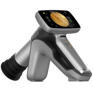
Zeiss Cirrus 4000 OCT — Refurbished Spectral-Domain OCT with RNFL, Macular & Ganglion Cell Normative Databases
The Zeiss Cirrus 4000 is a spectral-domain OCT (HD-OCT) system built around Zeiss’s 27,000 A-scan/second acquisition engine and proprietary FastTrac™ retinal tracking technology — delivering 5 µm axial resolution retinal imaging with macular cube, optic disc cube, and RNFL circle scan protocols that populate the Cirrus normative database for glaucoma and macular disease staging directly from each scan. This refurbished Zeiss Cirrus 4000 includes a Quad-Core processor, Windows 7, and table, configured as a turnkey OCT workstation ready for immediate clinical use in glaucoma monitoring, diabetic macular edema management, and AMD evaluation.

The Zeiss Cirrus 4000 captures 200×200 and 512×128 macular cube scans and 200×200 optic disc cube scans that provide the three-dimensional retinal dataset needed for ganglion cell analysis, RNFL deviation maps, and longitudinal progression tracking — all backed by normative databases built from hundreds of age-matched controls. Research published in Ophthalmology (National Library of Medicine, 2023) confirmed that peripapillary OCT scans are comparable to or better than standard automated perimetry for detecting glaucoma worsening — directly validating the RNFL-based glaucoma monitoring workflow that the Zeiss Cirrus 4000 is designed to perform in everyday clinical practice.
Why Practices Choose the Zeiss Cirrus 4000
- 27,000 A-scan/second acquisition — Zeiss’s HD-OCT engine captures the full macular cube (200×200: 40,000 A-scans) in under 2 seconds, minimizing motion artifact and enabling consistent image quality even in patients with fixation difficulties or media opacities.
- 5 µm axial resolution in tissue — Ultra-high axial resolution reveals individual retinal layer boundaries — the external limiting membrane, IS/OS junction, and RPE — that lower-resolution systems miss, enabling earlier detection of photoreceptor layer disruption in DME and AMD.
- RNFL + Macular + ONH + Ganglion Cell normative databases — Four separate normative databases with probability maps allow color-coded deviation displays for each measurement type, converting raw thickness numbers into clinically actionable normal/borderline/outside-normal-limits classifications on every scan.
- Ganglion Cell Analysis (GCA) — The Cirrus 4000 ganglion cell + inner plexiform layer (GCIPL) map targets the macular ganglion cells that represent 50% of all retinal ganglion cells, providing a second structural glaucoma endpoint independent of RNFL that detects loss in patients with confounding disc anatomy or RNFL artifacts.
- FastTrac™ retinal tracking — Real-time retinal motion tracking locks each A-scan to the same retinal location on every visit, ensuring that follow-up scans are registered to baseline for meaningful progression analysis rather than comparing different retinal locations across visits.
- Guided Progression Analysis (GPA) — The Cirrus GPA algorithm detects statistically significant RNFL change over time, flagging areas of possible or likely progression with color-coded maps that let the physician identify declining sectors before they cross below the normative database threshold.
- Anterior segment module compatibility — The Cirrus 4000 accepts the optional Anterior Segment Adapter for corneal pachymetry, anterior chamber angle imaging, and LASIK flap measurement — extending the instrument’s clinical utility beyond posterior segment imaging.
Clinical Applications
- Glaucoma diagnosis and progression monitoring — RNFL thickness maps with normative database color coding and the GCA module detect structural glaucomatous loss; GPA tracks progression over time to guide treatment escalation decisions in established glaucoma and glaucoma suspects.
- Diabetic macular edema (DME) assessment — Macular cube scans generate retinal thickness maps and ETDRS grid thickness measurements that quantify edema volume and document treatment response to anti-VEGF injections across serial visits.
- AMD monitoring and staging — High-resolution B-scans through the fovea characterize drusen morphology, RPE elevation, and subretinal/intraretinal fluid to stage dry AMD progression and document wet AMD treatment response.
- Macular disease documentation — The Cirrus 4000 documents epiretinal membranes, macular holes, vitreomacular traction, and cystoid macular edema with the cross-sectional B-scan detail needed to guide surgical decision-making in vitreoretinal referrals.
- Optic disc analysis — Disc cube scans measure cup-to-disc ratio, rim area, disc area, and cup volume with normative comparison — providing quantitative optic disc parameters that support glaucoma suspect management alongside RNFL analysis.
- Pre- and post-operative retinal assessment — OCT documents macular integrity before cataract surgery in patients with suspected macular pathology, and confirms photoreceptor layer restoration after retinal detachment repair or macular surgery.
Who This Is For
- Comprehensive ophthalmology and optometry practices adding OCT for the first time — the Cirrus 4000 normative databases and automated report generation make OCT interpretation accessible to any clinician without subspecialty fellowship training.
- Glaucoma-focused practices that need RNFL + GCA longitudinal monitoring — the Cirrus GPA algorithm tracks structural progression with statistical significance testing that supports documentation for managed care medical necessity reviews.
- High-volume cataract practices pre-screening surgical candidates — OCT confirms macular integrity in patients with potentially confounding pathology before committing to premium IOL recommendations.
- Retina co-management practices monitoring DME and AMD patients between subspecialty visits — the Cirrus 4000’s fluid detection and retinal thickness mapping provides the imaging documentation needed to triage urgent referrals from routine follow-up visits.
- Practices upgrading from time-domain OCT — the Zeiss Cirrus 4000’s 70× faster acquisition speed and 2× better resolution compared to first-generation OCT systems delivers visibly superior image quality and normative database analysis that time-domain systems cannot provide.
Technical Specifications
| Specification | Detail |
| OCT Technology | Spectral-Domain HD-OCT (SD-OCT) |
| A-Scan Rate | 27,000 Hz |
| Axial Resolution (tissue) | 5 µm |
| Transverse Resolution | 15 µm |
| Scan Protocols | Macular Cube 200×200, Macular Cube 512×128, Optic Disc Cube 200×200, HD 5-Line Raster, HD Single Line, RNFL and ONH OU Analysis |
| Analysis Modules | RNFL, Macular, ONH, Ganglion Cell Analysis (GCA), GPA Progression |
| Normative Databases | RNFL, Macular, ONH, Ganglion Cell + IPL |
| Motion Tracking | FastTrac™ real-time retinal tracking |
| Wavelength | 840 nm |
| Minimum Pupil | 2.5 mm |
| Processor | Quad-Core |
| Software Platform | Windows 7 |
| Manufacturer | Carl Zeiss Meditec, Germany |
| Condition | Refurbished |
| Warranty | 6 months |
Compare Zeiss Cirrus OCT Options
| Feature | Zeiss Cirrus 4000 | Zeiss Cirrus 4000 (Used) | Zeiss Cirrus 5000 |
| A-Scan Rate | 27,000 Hz | 27,000 Hz | 68,000 Hz |
| Ganglion Cell Analysis | Yes | Yes | Yes (enhanced) |
| GPA Progression | Yes | Yes | Yes |
| AngioPlex OCT-A | No | No | Yes |
| Condition | Refurbished | Used | Refurbished |
| Price | $13,900 | $15,400 | $26,000 |
Questions about the Zeiss Cirrus 4000? Contact us for availability and shipping details.