Description
This exact unit sold recently — but we specialize in sourcing refurbished ophthalmic equipment. Typical turnaround is 2–4 weeks once we confirm availability with our partners. Request a free quote with no deposit required.
Request Availability → Free quote · No obligation · 90-day warranty when shipped
What’s Included
- Zeiss Cirrus 5000 OCT with AngioPlex® OCTA
- Quad Core processor
- Windows 10 operating system
- Anterior segment module
- Adjustable exam table
- FastTrac™ retinal tracking
- 6-month Digital Eye Center warranty on parts and workmanship
- Optional: Premier Lens add-on (+$3,500)
Condition & Warranty
Refurbished. This Zeiss Cirrus 5000 with AngioPlex has been fully inspected and tested to factory performance standards. Covered by a 6-month warranty on parts and workmanship from Digital Eye Center.
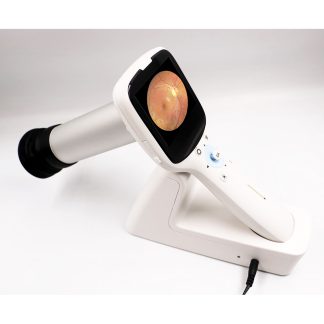
Zeiss Cirrus 5000 — Refurbished OCT with AngioPlex
 The Zeiss Cirrus 5000 is ZEISS’s mid-tier performance OCT platform combining 68,000 A-scans per second with integrated AngioPlex® OCT angiography for retina, glaucoma, and anterior segment in a single workflow. The Zeiss Cirrus 5000 delivers the same clinically validated FastTrac™ retinal tracking, Guided Progression Analysis™, and Ganglion Cell analysis as the Cirrus 6000 — at a price point accessible to mid-volume practices and growing clinics. This fully refurbished unit includes the anterior segment module, Windows 10, Quad Core processor, and adjustable table.
The National Library of Medicine confirms that OCT provides superior sensitivity for glaucoma screening compared to optic disc photography alone — the diagnostic advantage the Zeiss Cirrus 5000‘s RNFL and GCL/IPL analytics are specifically designed to deliver.
Why Practices Choose the Zeiss Cirrus 5000
- 68,000 A-scans/second with FastTrac™ — real-time retinal tracking registers every scan to prior visits, enabling true point-to-point macular and RNFL comparison across the patient record
- AngioPlex® OCTA included — 8×8, 6×6, and 3×3 mm OCTA scans with OMAG technology; non-invasive dye-free visualization of retinal microvasculature
- Smart HD scans — 19-inch HD display with enhanced B-scan detail; HD averaging reduces noise on challenging patients
- Guided Progression Analysis™ (GPA™) — ZEISS-exclusive trend and event analysis for RNFL, ONH, and GCL/IPL change detection, compatible with all Cirrus-generation data
- Ganglion Cell Analysis (GCL/IPL) — macular glaucoma detection that catches structural loss GPA may miss when peripapillary RNFL appears within normal limits
- Anterior segment module included — corneal topography and anterior segment OCT for surgical planning without additional hardware
- Full Cirrus data continuity — imports raw data from Cirrus 400 and 4000 platforms for uninterrupted longitudinal patient records
Clinical Applications
- Glaucoma detection and progression monitoring — RNFL thickness, GCL/IPL deviation maps, and GPA trend analysis from suspect through advanced; NDB comparison flags abnormal sectors automatically
- Diabetic retinopathy — AngioPlex OCTA quantifies FAZ, capillary dropout, and retinal perfusion changes across serial visits for anti-VEGF treatment monitoring
- Age-related macular degeneration — macular thickness change maps with automated serial comparison detect drusen progression and CNV activity; OCTA confirms neovascular AMD without dye
- Retinal vascular occlusions — OCTA maps perfusion deficits and macular ischemia for BRVO/CRVO assessment and treatment response
- Anterior segment imaging — corneal topography, epithelial mapping, and anterior segment OCT for surgical candidacy and post-op monitoring
- Post-treatment follow-up — retinal layer tracking after intravitreal injection, laser treatment, or vitreoretinal surgery using FastTrac™-registered serial scans
Who This Is For
- Comprehensive ophthalmology practices that need a full-featured OCTA platform for retina and glaucoma without the Cirrus 6000’s price premium
- Glaucoma specialists prioritizing GPA progression analysis, GCL/IPL mapping, and RNFL trending across a large patient panel
- Retina-focused practices managing diabetic retinopathy and AMD with regular OCTA follow-up visits
- Practices upgrading from Cirrus 400 or 4000 — existing patient data imports automatically; staff training is minimal
- Mid-volume clinics where the Cirrus 6000’s 100K scan speed is not a bottleneck but full AngioPlex capability is required
Technical Specifications
| Specification | Detail |
| Scan Speed | 68,000 A-scans/second |
| Axial Resolution | 5 µm (tissue) |
| Transverse Resolution | 15 µm (tissue) |
| OCTA Module | AngioPlex® (8×8, 6×6, 3×3 mm) |
| Eye Tracking | FastTrac™ real-time retinal tracking |
| Display | 19-inch HD |
| Processor | Quad Core |
| Anterior Segment | Included (Premier lens optional +$3,500) |
| Wavelength | 840 nm SLD |
| Operating System | Windows 10 |
| Condition | Refurbished |
| Warranty | 6 months |
Compare Zeiss Cirrus 5000 with Other OCT Options
| Feature | Zeiss Cirrus 5000 + AngioPlex | Zeiss Cirrus 5000 (no OCTA) | Zeiss Cirrus 4000 |
| Scan Speed | 68,000/sec | 68,000/sec | 27,000/sec |
| AngioPlex OCTA | Included | Not included | Not available |
| Max OCTA Field | 8×8 mm | — | — |
| FastTrac™ Tracking | Yes | Yes | Yes |
| GPA Analysis | Yes | Yes | Yes |
| Price | $35,200 | $26,000 | $15,400 |
Also consider the Zeiss Cirrus 5000 without AngioPlex for structural OCT at a lower price, or the Zeiss Cirrus 4000 as an entry-level option.
Similar OCTs In Stock Now
Need a comparable oct available today? These units are in stock and ready to ship with a 90-day Digital Eye Center warranty.


Prefer to wait for the Zeiss Cirrus 5000A OCT w/ Angioplex? Request availability and we’ll source one for you.
Questions about the Zeiss Cirrus 5000? Contact us for availability and shipping details.