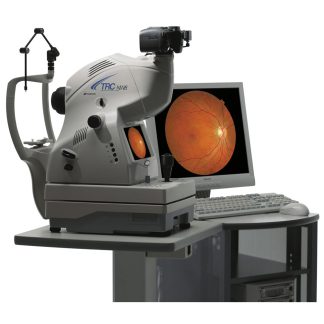
Description
Specifications of the Visucam Pro NM/FA Fundus camera – Non-Mydriatic
VISUCAM NM/FA With advanced imaging options for autofluorescence and ICG angiography
Autofluorescence
VISUCAM® NM/FA is now available with an autofluorescence option, for easy and fast monitoring of AMD patients. Exposure time is reduced, which means the VISUCAM® NM/FA is available for use with other patients faster and patient stress is minimized..
ICG angiography
For clinicians who want to perform occasional ICGA procedures, the ICG angiography option allows you to compare fluorescein angiography, autofluorescence and indocyanin angiography directly at the VISUCAM® NM/FA. This comprehensive analysis allows immediate and precise diagnoses and also reduces the procedure time.
Auto focus
Auto focus enables a smooth and fast workflow while reducing your operating steps.
Small pupil capabilities
Non-mydriatic image capture of fundus imaging through the smallest pupil size in the industry.
All-in-one approach
High-quality ZEISS fundus imaging in a compact system:
- Highly corrected ZEISS optics with an advanced professional-grade digital sensor
- Integrated computer and database, including multiple options for image comparison and review
- Quick image transfer via network, USB stick, or DVD.

Image is Everything.
ZEISS optics
The demonstrated quality of ZEISS optics assures that your image will give you the sharpest detail.
5.0 megapixels images
The pixel count and optimized signal processing enable outstanding quality in image documentation.
45° and 30° images
The system features the ophthalmoscope principle incorporated in modern fundus cameras and two field angles: 45° and 30°.
Different capture modes
Green, red, and blue images are all possible as separate photos or as a subsequent RGB layer of a color photo.
Stereo image mode
The stereo mode features easy capture and operation.
Fixation mode
A single click suffices to switch between external and internal fixation. The internal targets are either freely positionable in different sizes or follow programmed sequences.
Zeiss Automap
Structured montages are automatically produced of peripheral photos.

Optimized practice efficiency.
Everything is integrated
The Zeiss Visucam Pro NM offers the entire performance spectrum—from image capture to image documentation—in a single, state-of-the-art system featuring all required hardware and software.
Everything is simple
Easy operation ensures a smooth, rapid workflow for the user. The system’s many excellent benefits include:
• Customized software settings
• Positioning aid with working distance dots
• Focusing aid with paired coincidence lines
• Panorama mode for automatic image montages
Everything is visible
Visual overview and assessment are always possible in every examination phase – thanks to the 17″ flat screen monitor. The path from the live focusing image to the digital document is short. When captured, the image immediately appears on the monitor and is automatically stored.
Everything is possible
With its excellent image quality, Visucam Pro NM is the perfect solution for studies. The 7-field mode, the 30° field angle, and 3D images are integrated for this purpose.
Everything is reliable
Software manages image display, editing, printing and data export. Password-protected user management pre- vents unauthorized access to images and patient data. VISUPAC performs archiving in networks, while DVDs/CDs or USB sticks are more suitable for simple export.

Specifications for Zeiss Visucam Pro NM
| Image capture | |
| Field angle | 45° and 30° |
| Capture modes | Color, red-free, blue and red pictures and pictures of the anterior segment, as well as fluorescein angiography |
| Filters | Blue and red-free filters, UV and IR barrier filters |
| Capture sequence | 1.5 s – 2.5 s (depends on flash energy) |
| Compensation for ametropia | –35 D … +40 D |
| Pupil diameter | ≥ 3.3 mm (WF small pupil mode) |
| Working distance | 40 mm (patient’s eye – front lens) |
| Capture sensor | CCD 5 megapixels |
| Monitor | 19” TFT (1280 × 1024 pixels) |
| Fixation | |
| External and internal | Vertically programmed sequences or freely positionable |
| Flash energy | Xenon flash lamp, 22 flash levels |
| Database | Patient information and images with field angle, date, time, description and side of view |
| Computer | |
| Operating system | Windows XP Professional Use of other operating systems on request |
| Hard disk | 160 GB, minimum 60,000 images |
| Export/import | JPEG, BMP, TIFF, DICOM, Stimons, png |
| Printer | INKJET, B/W, color |
| Internal DVD burner | R/W format: DVD, CD |
| Dimensions | |
| Instrument | (W × D × H) 480 × 635 × 546 mm (18.9 × 25 × 21.5 in.) |
| Weight of basic system | Approx. 35 kg (77.2 lbs) |
| Line voltage | 100 – 240 V ± 10 % (self-adjusting) |
| Frequency | 50/60 Hz |
| Instrument table | Asymmetric, suitable for wheelchair |
| Accessories | USB memory stick VISUPAC archiving and image analysis system Network adapter |