Slit lamp photography has gone from a niche capability reserved for academic medical centers to a practical, affordable standard for any eye care practice. Whether you see ten patients a day or a hundred, documenting what you observe through the slit lamp — corneal scars, anterior chamber findings, lens changes, filtering blebs — protects your patients, supports your documentation, and makes clinical communication dramatically clearer.
This guide covers why slit lamp photography matters, what equipment makes it possible, and how practices of every size can get started without overcomplicating it.
Why Slit Lamp Photography Has Become Essential
The slit lamp remains the most powerful diagnostic tool in an eye care practice — but until recently, everything seen through it stayed in the examiner’s head and a handwritten note. That gap matters more today than ever. Patients expect to see and understand their findings. Insurance carriers require photographic documentation for many anterior segment conditions. Telemedicine referrals are meaningless without images. And for tracking progressive conditions like pterygium, corneal dystrophies, or post-surgical changes over months and years, nothing beats a dated photograph. A study published in Optometry and Vision Science (National Library of Medicine) confirmed that digital photography through the slit lamp provides clinically valid anterior segment measurements — supporting its use not just for records, but as a genuine diagnostic tool.
What You Need for Slit Lamp Photography
The good news is that you don’t need a new slit lamp. The slit lamp you already own — whether it’s a Haag-Streit, Topcon, Zeiss, or any standard model — can be outfitted with a camera adapter that attaches to the existing eyepiece or beam splitter port. The equipment falls into three tiers based on volume and workflow needs.
Tier 1 — Smartphone Adapters
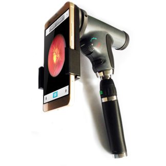
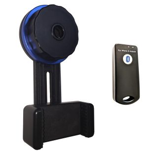
A universal smartphone slit lamp adapter clamps your iPhone or Android phone over the eyepiece and lets you capture anterior segment images directly to your device. The images transfer instantly to your EMR or can be shared with patients on the spot. The Universal Slit Lamp Adapter with Bluetooth Remote eliminates camera shake at the moment of capture — a common reason smartphone slit lamp photos come out blurry. This is the lowest-cost entry point and the right choice for practices doing occasional documentation or running mobile screening programs.
Tier 2 — Dedicated Camera Adapters
A dedicated camera mounted on the slit lamp via a C-mount or DSLR adapter stays in place full-time and is always ready to shoot — no phone to attach, no case to remove. Images record to the camera’s memory or transfer wirelessly to a networked PC. Digital Eye Center carries several options, including the Microscope Camera Adapter C-Mount Full HD with a compact camera included, the Slit Lamp Camera Adapter for Canon / Nikon DSLR for practices that own a compatible body, and a C-mount video camera adapter for standard industry cameras. These are the practical sweet spot for practices that want clinical-grade images without the complexity of a full beam splitter system.
Tier 3 — Beam Splitter Systems
A beam splitter divides the slit lamp’s optical path so that the examiner and the camera view the same field simultaneously — the camera runs continuously in the background without interrupting the exam. This is the standard in high-volume clinics and teaching settings. Beam splitter sets are slit lamp-specific: the Haag-Streit Beam Splitter Set for BQ900 / BX900, the Topcon / Marco Beam Splitter Set, and the Zeiss C-Mount Adapter Set are all available and ship from our Florida warehouse.
Tips for Better Slit Lamp Photos
- Use the lowest magnification that shows the finding. Higher magnification narrows the field and makes focus more critical — start wide, then zoom in.
- Dim the room lights. Ambient light washes out the slit beam and reduces contrast in the image.
- Use a Bluetooth or wired remote shutter. Pressing the camera button directly — even gently — introduces enough vibration to blur fine corneal detail.
- Document every significant finding at baseline. A photo taken today is useless without something to compare it to six months from now.
- Label your images at the time of capture. Most dedicated cameras and EMRs support date-stamped file naming — use it consistently so images are findable later.
Shop Slit Lamp Photography Equipment
- Universal Slit Lamp Smartphone Adapter w/ Bluetooth Remote
- Microscope Camera Adapter C-Mount Full HD
- Slit Lamp Camera Adapter for Canon / Nikon DSLR
- Haag-Streit Beam Splitter Set (BQ900 / BX900)
- Topcon / Marco Beam Splitter Set
- Zeiss C-Mount Adapter Set
Contact us for compatibility questions, availability, and shipping details.